By Peplyte 27 days ago
ALL ARTICLES AND PRODUCT INFORMATION PROVIDED ON THIS WEBSITE ARE SOLELY FOR INFORMATION DISSEMINATION AND EDUCATIONAL PURPOSES.
The products provided on this website are intended exclusively for in vitro research. In vitro research (Latin: *in glass*, meaning in glassware) is conducted outside the human body. These products are not pharmaceuticals, have not been approved by the U.S. Food and Drug Administration (FDA), and must not be used to prevent, treat, or cure any medical condition, disease, or ailment. It is strictly prohibited by law to introduce these products into the human or animal body in any form.
Overview
HIV infection and antiretroviral therapy (ART)-induced fat metabolism abnormalities pose serious challenges to the health and quality of life of HIV patients. These abnormalities manifest in various forms, such as lipoatrophy and lipohypertrophy, with excessive accumulation of visceral adipose tissue (VAT) being particularly prominent. The increase in VAT not only affects patients’ physical appearance but is also closely associated with a range of severe metabolic complications, including cardiovascular disease, insulin resistance, and non-alcoholic fatty liver disease (NAFLD), posing a serious threat to patients’ long-term survival and health status.
Tesamoreli, a synthetic growth hormone-releasing hormone (GHRH) analog, offers new hope for the treatment of fat metabolism abnormalities in HIV patients. It specifically binds to GHRH receptors in the anterior pituitary gland, effectively stimulating the synthesis and release of endogenous growth hormone (GH). GH plays a crucial role in regulating fat metabolism in the human body, promoting lipolysis, enhancing fatty acid oxidation, and regulating the expression of genes related to lipid metabolism, thereby exerting a positive influence on fat metabolism. This unique mechanism of action enables Tesamoreli to help improve fat metabolism disorders in HIV patients.
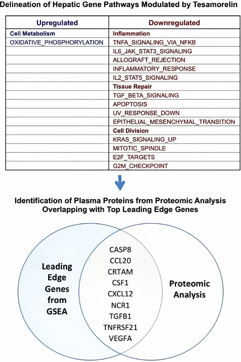
Figure 1 Analysis Schema. A total of 9 plasma proteins were examined, corresponding to top leading edge genes within hepatic gene pathways differentially modulated by tesamorelin.
Mechanism of Action of Tesamoreli on Fat Metabolism in HIV
Patients
Regulation of the Growth Hormone-Insulin-Like Growth Factor Axis
Tesamoreli binds to the GHRH receptor on the cell membrane of the anterior pituitary gland, activating receptor-associated signaling pathways such as the JAK-STAT signaling pathway. This activation process promotes the synthesis and release of growth hormone (GH) by the anterior pituitary gland. After entering the bloodstream, reaches target organs such as the liver, stimulating the liver to synthesize and secrete insulin-like growth factor-1 (IGF-1). IGF-1 serves as a key mediator of GH’s metabolic effects and plays a crucial role in fat metabolism. IGF-1 can activate the PI3K-AKT signaling pathway, inhibit adipocyte apoptosis, and maintain normal adipocyte function. It also promotes the uptake and oxidation of fatty acids by adipocytes, reducing fat accumulation within cells. In HIV patients, the function of the growth hormone-insulin-like growth factor axis is often impaired due to the disease itself and the effects of antiretroviral therapy (ART). Tesamoreli can activate this axis to restore its normal fat metabolism regulatory function.
Direct effects on adipocyte metabolism
Promoting lipolysis: Tesamoreli can upregulate the expression and activity of hormone-sensitive lipase (HSL) in adipocytes. HSL is a key enzyme in lipolysis, catalyzing the hydrolysis of triglycerides into glycerol and fatty acids, thereby promoting the catabolic metabolism of fat. In vitro, adding Tesamoreli to cultured adipocytes significantly enhances HSL activity and markedly increases fatty acid release. Tesamoreli can also indirectly activate HSL by regulating the cyclic AMP (cAMP)-protein kinase A (PKA) signaling pathway, further promoting lipolysis. In HIV patients, this lipolysis-promoting effect helps reduce excessive fat accumulation, particularly visceral fat reduction, thereby improving abnormal fat distribution in patients.
Regulation of fatty acid oxidation: Tesamoreli enhances the activity of key enzymes involved in fatty acid oxidation within adipocytes, such as carnitine palmitoyltransferase 1 (CPT1). CPT1 is the rate-limiting enzyme for fatty acid entry into mitochondria for β-oxidation, and its enhanced activity promotes the oxidative breakdown of fatty acids within mitochondria, providing energy for cells. Tesamoreli can also upregulate the expression of other enzymes and transporters involved in fatty acid β-oxidation, such as acetyl-CoA carboxylase 2 (ACC2) and fatty acid transporter 1 (FATP1), further optimizing the fatty acid oxidation metabolic process. By promoting fatty acid oxidation, Tesamoreli can reduce fat storage within cells and improve lipid metabolism disorders in HIV patients.
Regulation of lipid metabolism-related gene expression
Influencing adipogenesis-related genes: Tesamoreli can inhibit the expression of adipogenesis-related genes, such as peroxisome proliferator-activated receptor γ (PPARγ) and CCAAT/enhancer-binding protein α (C/EBPα). PPARγ is a key transcription factor in adipocyte differentiation and adipogenesis, promoting the differentiation of fibroblasts into mature adipocytes, and induces the expression of a series of adipogenesis-related genes, such as fatty acid-binding protein 4 (FABP4) and fatty acid synthase (FAS). C/EBPα also plays a crucial role in adipocyte differentiation and adipogenesis. By inhibiting the expression of PPARγ and C/EBPα, Tesamoreli reduces adipocyte differentiation and adipogenesis, thereby decreasing fat accumulation in the body. In HIV patients, this regulatory effect on adipogenesis-related genes helps correct the excessive fat production caused by abnormal fat metabolism.
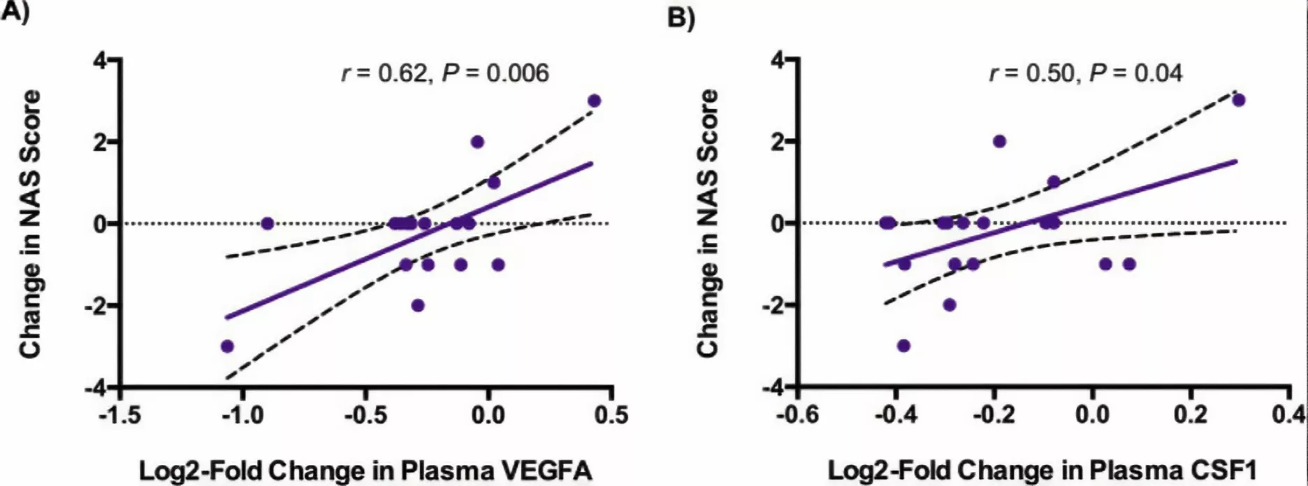
Figure 2 Relationship of Changes in Plasma VEGFA and CSF1 with Change in NAS Score in Tesamoreli-Treated Participants.
Regulation of lipid transport and metabolism genes: Tesamoreli can regulate the expression of genes related to lipid transport and metabolism, such as members of the apolipoprotein (Apo) family. ApoB is the primary apolipoprotein of very-low-density lipoprotein (VLDL) and low-density lipoprotein (LDL), and its expression levels are closely correlated with the levels of VLDL and LDL in plasma. Tesamoreli can reduce ApoB expression, reduce the synthesis and secretion of VLDL and LDL, thereby lowering plasma levels of atherogenic lipoproteins. Additionally, Tesamoreli can upregulate the expression of ApoA-I, the primary apolipoprotein of high-density lipoprotein (HDL). Increased ApoA-I expression helps elevate HDL levels, promote cholesterol reverse transport, and reduce the risk of cardiovascular disease. In HIV patients, where lipid metabolism disorders often accompany lipid metabolism abnormalities, Tesamoreli’s regulatory effects on lipid transport and metabolism genes can help improve patients’ lipid profiles.
Specific Effects of Tesamoreli on Lipid Metabolism in HIV Patients
Reducing Visceral Adipose Tissue (VAT)
Tesamoreli significantly reduces VAT in HIV patients. In a Phase III clinical trial involving HIV patients, participants were randomly assigned to receive either 2 mg of Tesamoreli daily or a placebo for 26 weeks. In the Tesamoreli treatment group, patients exhibited a significant reduction in VAT, and this reduction was observed across patients with different baseline characteristics (e.g., presence or absence of back-neck fat). For Tesamoreli responders (defined as patients with at least an 8% reduction in VAT and who adhered to treatment), VAT decreased significantly regardless of the presence of neck and back fat, and there was no statistically significant difference between the two groups (P = 0.657). This suggests that Tesamoreli has broad efficacy in reducing VAT in HIV patients, independent of the presence of back-neck fat. The reduction in VAT not only improves patients’ physical appearance but more importantly reduces the risk of VAT-related metabolic complications, such as cardiovascular disease and insulin resistance.
Improving liver fat metabolism
Reducing liver fat content: In HIV patients, the incidence of non-alcoholic fatty liver disease (NAFLD) is high and closely associated with abnormal fat metabolism. Tesamoreli has a positive therapeutic effect on HIV-related NAFLD. In a randomized, double-blind, multicenter study, HIV-infected patients with a hepatic fat fraction (HFF) ≥5% were randomly assigned to the tesamorelin group or the placebo group and treated for 12 months. The results showed that patients in the Tesamoreli group had a more significant reduction in HFF, with an absolute effect size of -4.1% (95% CI -7.6 to -0.7, P = 0.018) and a relative reduction of 37% from baseline (95% CI -67 to -7, P = 0.016). After 12 months of treatment, 35% of patients in the Tesamoreli group had HFF levels below 5%, compared to only 4% in the placebo group (P = 0.0069). This indicates that Tesamoreli effectively reduces liver fat content and improves liver fat metabolism in HIV patients.
Potential effects on liver histology: Analysis of liver gene expression profiles in HIV-NAFLD patients revealed that Tesamoreli downregulated gene sets associated with inflammation, tissue repair, and cell division in the liver. Further proteomics studies showed that Tesamoreli led to a significant reduction in plasma protein levels of vascular endothelial growth factor A (VEGFA), transforming growth factor β1 (TGFB1), and macrophage colony-stimulating factor 1 (CSF1), which are closely associated with liver fibrosis and inflammation. In patients treated with Tesamoreli, the reduction in plasma VEGFA and CSF1 was associated with a decrease in NAFLD activity scores, while the reduction in TGFB1 and CSF1 was associated with a decrease in gene-level fibrosis scores. This suggests that Tesamoreli may exert beneficial effects on liver histology by regulating inflammation and fibrosis-related pathways in the liver.
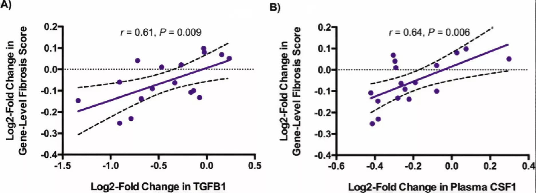
Figure 3 Relationship of Changes in Plasma TGFB1 and CSF1 with Change in Gene-Level Fibrosis Score in Tesamoreli-Treated Participants.
Regulation of Whole-Body Fat Distribution and Body Composition
Reducing trunk fat: In addition to reducing VAT, Tesamoreli also has a significant regulatory effect on trunk fat in HIV patients. In clinical studies, patients treated with Tesamoreli showed a gradual reduction in trunk fat content, which helps improve body shape and reduce issues such as abdominal protrusion caused by abnormal fat distribution. The reduction in trunk fat is interrelated with the reduction in VAT, collectively improving body composition.
Improving waist circumference: Waist circumference is an important indicator of abdominal fat accumulation. Tesamoreli treatment significantly improves waist circumference in HIV patients. In relevant clinical trials, both Tesamoreli responders with and without back-neck fat showed a significant reduction in waist circumference after 26 weeks of treatment. The improvement in waist circumference not only reflects reduced abdominal fat but is also closely associated with reduced metabolic risks such as cardiovascular disease.
Effects on other body composition parameters: Tesamoreli also has a regulatory effect on other body composition parameters in HIV patients. For example, in some studies, while body mass index (BMI) showed no significant changes, body fat composition improved. Tesamoreli can optimize body fat distribution without affecting overall weight, reduce excessive fat accumulation, increase lean body mass components such as muscle, and enhance patients’ body quality.
Application of Tesamoreli in Fat Metabolism in HIV Patients
Target Population
HIV patients with abnormal fat metabolism: Tesamoreli is primarily indicated for HIV patients with abnormal fat metabolism, particularly those with excessive visceral fat (e.g., as determined by abdominal CT or MRI scans showing visceral adipose tissue [VAT] exceeding normal ranges). These patients typically exhibit abdominal obesity, increased waist circumference, and may also present with other manifestations of metabolic syndrome, such as insulin resistance and dyslipidemia.
Conclusion
As an effective therapeutic agent for abnormal fat metabolism in HIV patients, Tesamoreli regulates fat metabolism through multiple mechanisms, playing a significant role in reducing VAT, improving hepatic fat metabolism, regulating whole-body fat distribution, and optimizing body composition.
Sources
[1] Rahman F, de Chantal M, Mesquita P, et al. 935. Effect of Tesamoreli in People with HIV with and without Dorsocervical Fat: Post Hoc Analysis of Phase III Double Blind Placebo Control Trial[J]. Open Forum Infectious Diseases, 2020,7(Supplement_1):S500-S501.DOI:10.1093/ofid/ofaa439.1121.
[2] Clinical Review Report: Tesamoreli (Egrifta)[M]. Ottawa (ON): Canadian Agency for Drugs and Technologies in Health, 2016.
http://www.ncbi.nlm.nih.gov/entrez/query.fcgi?cmd=Retrieve&db=pubmed&dopt=Abstract&list_uids=30920787&query_hl=1.
Product available for research use only:
<
By Peplyte 29 days ago
ALL ARTICLES AND PRODUCT INFORMATION PROVIDED ON THIS WEBSITE ARE SOLELY FOR INFORMATION DISSEMINATION AND EDUCATIONAL PURPOSES.
The products provided on this website are intended exclusively for in vitro research. In vitro research (Latin: *in glass*, meaning in glassware) is conducted outside the human body. These products are not pharmaceuticals, have not been approved by the U.S. Food and Drug Administration (FDA), and must not be used to prevent, treat, or cure any medical condition, disease, or ailment. It is strictly prohibited by law to introduce these products into the human or animal body in any form.
Overview
Respiratory health has long been a focal point of research in the fields of medicine and life sciences. As our understanding of the mysteries of life deepens, the roles of various bioactive substances in maintaining normal respiratory physiological functions and in the onset and progression of diseases have gradually been elucidated. The association between Bronchogen and respiratory health not only involves fundamental physiological processes but is also closely linked to the pathological mechanisms of various respiratory diseases.

Biological Characteristics of Bronchogen
(1) Structural Characteristics
Bronchogen possesses a unique molecular structure. It contains specific functional domains that participate in interactions with other biomolecules, such as binding to cell membrane surface receptors and recognizing proteins associated with intracellular signaling pathways. This structural framework forms the basis for its biological functions, determining its target sites and modes of action within the respiratory tract.
(2) Sources and Distribution
Within the body, Bronchogen has a wide range of sources. It can be synthesized and secreted by local cells in the respiratory tract, such as respiratory epithelial cells and immune cells. These cells activate the expression of relevant genes in response to specific stimuli, thereby synthesizing Bronchogen. Bronchogen may also be transported to the respiratory tract via the bloodstream from other tissues and organs. In terms of distribution, Bronchogen is present at certain concentrations throughout the respiratory tract, including the nasal cavity, pharynx, trachea, bronchi, and alveoli. The concentration of Bronchogen may vary across different regions due to local physiological functions and pathological states. This distribution pattern is closely related to the physiological functional zones of the respiratory tract and the regions where diseases commonly occur.
The Role of Bronchogen in Respiratory Physiological Functions
(1) Immunomodulatory Effects
Regulation of Immune Cell Activity
Bronchogen can finely regulate the activity of immune cells within the respiratory tract. It can enhance the phagocytic function of macrophages, thereby strengthening their ability to recognize and eliminate pathogens. As a key defense line of the respiratory tract immune system, enhanced macrophage function facilitates the rapid clearance of invading bacteria, viruses, and other pathogens, thereby maintaining immune balance in the respiratory tract. Bronchogen also regulates the differentiation and proliferation of T lymphocytes and B lymphocytes, influencing the intensity and direction of specific immune responses. When facing pathogen infections, Bronchogen can guide T lymphocytes to differentiate into different subtypes such as Th1 or Th2, thereby determining whether the immune response is cell-mediated or humoral-mediated, ensuring that the immune system can adopt the most effective response strategy based on the type of pathogen.
Regulation of immune factor secretion
During immune regulation, Bronchogen also plays a key regulatory role in the secretion of various immune factors. It can promote the moderate secretion of pro-inflammatory factors such as interleukins (e.g., IL-1, IL-6) and tumor necrosis factor (TNF-α). These pro-inflammatory factors recruit immune cells to the infection site during the early stages of infection, initiating an inflammatory response, to combat pathogen invasion. Bronchogen also prevents the excessive secretion of these pro-inflammatory factors, avoiding uncontrolled inflammatory responses that could damage respiratory tract tissues. Additionally, Bronchogen promotes the secretion of anti-inflammatory factors (such as IL-10), which exert anti-inflammatory effects during the later stages of inflammation, facilitating the repair of respiratory tract tissues and the resolution of inflammation, thereby maintaining the stability of the respiratory tract’s internal environment.
(2) Maintaining the Integrity of Respiratory Epithelial Cells
Promoting cell proliferation and repair
Respiratory epithelial cells serve as the first physical barrier of the respiratory tract, and their integrity is crucial for defending against pathogen invasion. Bronchogen promotes the proliferation of respiratory epithelial cells and accelerates the repair of damaged epithelial cells. Following respiratory tract injury caused by external stimuli (such as smoke or chemicals), Bronchogen activates intracellular signaling pathways, including the MAPK signaling pathway and the PI3K-Akt signaling pathway. Activation of these pathways promotes cell cycle progression, enabling epithelial cells to transition from the quiescent phase to the proliferative phase, thereby increasing cell numbers to fill damaged areas and restore epithelial cell integrity.
Regulation of intercellular connections
In addition to promoting cell proliferation, Bronchogen also participates in regulating the connections between respiratory epithelial cells. Epithelial cells form a continuous barrier through structures such as tight junctions and adherens junctions. Bronchogen can regulate the expression and distribution of these junctional proteins (such as occludin and claudin), maintaining the stability of intercellular connections. When respiratory epithelial cells are infected by pathogens or stimulated by inflammation, intercellular junctions may be disrupted, leading to impaired barrier function. Bronchogen can promptly repair and strengthen these junctions, preventing pathogens and harmful substances from penetrating the epithelial cell layer into the respiratory tissue, thereby ensuring the normal physiological function of the respiratory tract.
(3) Regulation of airway mucus secretion and clearance
Mucus secretion regulation
Airway mucus is an essential component of the respiratory tract’s defense system, capable of capturing and clearing inhaled pathogens, dust, and other foreign particles. Bronchogen plays a regulatory role in airway mucus secretion. It achieves this by binding to receptors on the surface of respiratory epithelial cells, activating intracellular signaling pathways, and regulating the expression of mucin genes and the synthesis and secretion of mucus in mucus-secreting cells (such as goblet cells). Appropriate mucus secretion is crucial for maintaining respiratory tract hydration and normal defensive functions. Bronchogen precisely regulates mucus secretion levels according to the physiological needs of the respiratory tract, ensuring that mucus effectively captures foreign particles without causing airway obstruction due to excessive secretion.
Mucus Clearance Promotion
In addition to regulating mucus secretion, Bronchogen also promotes mucus clearance in the airways. It enhances the frequency and amplitude of ciliary beating in the respiratory tract. As the “sweepers” of the respiratory tract, cilia’s rhythmic beating propels mucus and the foreign particles it carries toward the airway opening, which is then expelled from the body through coughing or other means. Bronchogen enhances ciliary motility by influencing ion channels and signaling pathways within ciliated cells, such as regulating calcium ion concentration and activating protein kinases, thereby improving mucus clearance efficiency and maintaining airway patency.
The Association Between Bronchogen and Respiratory Diseases
(1) Asthma
Changes in Bronchogen Levels in Asthma Patients
In asthma patients, the levels and functions of Bronchogen often undergo significant changes. Studies have found that during asthma attacks, the concentration of Bronchogen in the airways may abnormally increase or decrease. In some severe asthma patients, the levels of Bronchogen in airway secretions are significantly lower than in healthy individuals, which may be related to damage to airway epithelial cells, leading to reduced synthesis and secretion of Bronchogen. In some mild asthma patients, although Bronchogen concentrations in the airways may remain within normal ranges, functional assessments reveal reduced capacity to regulate immune cell activity and anti-inflammatory factor secretion, suggesting altered biological activity of Bronchogen in asthma patients.
The Role of Bronchogen in the Pathogenesis of Asthma
From the perspective of asthma pathogenesis, Bronchogen is involved in multiple pathways. Abnormal regulation of immune function by Bronchogen leads to excessive immune responses to allergens in asthma patients. In asthma patients, Bronchogen dysfunction results in excessive activation of Th2 cells, which secrete large amounts of cytokines (such as IL-4, IL-5, IL-13, etc.), These cytokines stimulate the infiltration and activation of inflammatory cells such as eosinophils in the airways, triggering airway inflammation and airway hyperresponsiveness. The weakened role of Bronchogen in maintaining the integrity of respiratory epithelial cells makes these cells more susceptible to damage from allergens and inflammatory mediators, further exacerbating airway inflammation and airway remodeling, thereby promoting the onset and progression of asthma.
(2) Chronic Obstructive Pulmonary Disease (COPD)
Characteristics of Bronchogen in COPD Patients
In the airways of COPD patients, Bronchogen also exhibits characteristics distinct from those in healthy individuals. As COPD progresses, the concentration and distribution of Bronchogen in the airways undergo changes. In the lung tissue and airway secretions of COPD patients, the concentration of Bronchogen may gradually decrease, and its expression in airway epithelial cells and immune cells is significantly reduced. These changes are closely associated with the decline in lung function and the exacerbation of airway inflammation in COPD patients. The molecular structure of Bronchogen in COPD patients may undergo modifications, leading to reduced biological activity and further impairing its normal function in the respiratory tract.
The role of Bronchogen in the pathological process of COPD
Bronchogen also plays an important role in the pathological process of COPD. The main pathological features of COPD include airway inflammation, excessive mucus secretion, destruction of lung parenchyma, and airway remodeling. Due to the weakened regulation of immune function by Bronchogen, inflammatory responses in the airways persist and are difficult to control. Inflammatory cells such as neutrophils and macrophages accumulate in large numbers in the airways, releasing various inflammatory mediators and proteases, leading to lung tissue damage and airway structural destruction. The imbalance in Bronchogen’s regulation of airway mucus secretion and clearance results in excessive mucus production and impaired clearance, further exacerbating airway obstruction. The reduced ability of Bronchogen to promote respiratory epithelial cell repair and maintain intercellular connections accelerates the process of airway remodeling, ultimately leading to progressive deterioration of lung function in COPD patients.
(3) Respiratory Infectious Diseases
The Role of Bronchogen in Bacterial Infections
During respiratory bacterial infections, Bronchogen participates in the body’s immune defense process. When bacteria invade the respiratory tract, Bronchogen can activate immune cells, enhancing their phagocytic and cytotoxic capabilities against bacteria. Bronchogen also regulates the secretion of immune factors, creating an immune microenvironment conducive to antibacterial activity. It can promote the expression of antimicrobial peptides, which directly act on bacteria, disrupting their cell membranes and cell walls, thereby inhibiting bacterial growth and reproduction. However, in some severe bacterial infections, pathogens may interfere with Bronchogen synthesis and function, leading to a decline in the body’s immune defense capabilities and making the infection difficult to control.
The Role of Bronchogen in Viral Infections
Bronchogen also plays a crucial role in respiratory viral infections. During the early stages of viral infection, Bronchogen can activate innate immune cells (such as dendritic cells and natural killer cells) to initiate antiviral immune responses. It promotes the secretion of antiviral cytokines such as interferon, which can inhibit viral replication and spread, limiting viral dissemination within the respiratory tract. Bronchogen also regulates adaptive immune responses, promoting the recognition and response of T lymphocytes and B lymphocytes to viral antigens, leading to the production of specific antibodies and cytotoxic T cells, thereby effectively eliminating virus-infected cells.
Conclusion
Bronchogen plays a crucial role in maintaining respiratory health and in the progression of respiratory diseases. In various respiratory conditions such as asthma, COPD, and respiratory infectious diseases, the levels, functions, and distribution of Bronchogen are altered, and it is involved in the pathophysiological mechanisms of these diseases.
Sources
[1] Basha L, Hamze M, Socarras A, et al. Respiratory health and the Syrian conflict: a scoping literature review[J]. Med Confl Surviv, 2024,40(2):111-152.DOI:10.1080/13623699.2024.2343996.
[2] Herrero-Cortina B, Lee A L, Oliveira A, et al. European Respiratory Society statement on airway clearance techniques in adults with bronchiectasis[J]. European Respiratory Journal, 2023,62(1).DOI:10.1183/13993003.02053-2022.
[3] Miller M D. Citing Foreign-Language Resources and Translations[M]//Miller M D. Discovering Hidden Gems in Foreign Languages. Cham: Springer International Publishing, 2023:251-265.DOI: 10.1007/978-3-031-18479-6_7.
[4] Solomen S, Aaron P. Techniques in Cardiopulmonary physiotherapy[M]. 2017. ISBN: 9788184452334
[5] Agrawal A, Mabalirajan U. Rejuvenating cellular respiration for optimizing respiratory function: targeting mitochondria[J]. American Journal of Physiology-Lung Cellular and Molecular Physiology, 2016,310(2):L103-L113.DOI:10.1152/ajplung.00320.2015.
[6] Durieux R, Lavigne J P, Scagnol I, et al. Intrapericardial bronchogenic cyst adherent to the ascending aorta[J]. Thoracic and Cardiovascular Surgeon, 2014,62(2):189-191.DOI:10.1055/s-0031-1298060.
[7] Monaselidze J R, Khavinson V, Gorgoshidze M Z, et al. Effect of the peptide bronchogen (Ala-Asp-Glu-Leu) on DNA thermostability[J]. Bulletin of Experimental Biology and Medicine, 2011,150(3):375-377.DOI:10.1007/s10517-011-1146-x.
Product available for research use only:
By Peptide Information May 1, 2025
ALL ARTICLES AND PRODUCT INFORMATION PROVIDED ON THIS WEBSITE ARE SOLELY FOR INFORMATION DISSEMINATION AND EDUCATIONAL PURPOSES.
The products provided on this website are intended exclusively for in vitro research. In vitro research (Latin: *in glass*, meaning in glassware) is conducted outside the human body. These products are not pharmaceuticals, have not been approved by the U.S. Food and Drug Administration (FDA), and must not be used to prevent, treat, or cure any medical condition, disease, or ailment. It is strictly prohibited by law to introduce these products into the human or animal body in any form.
Polypeptide Purity Control and Verification Technical System
peplyte.com provides peptides with purity exceeding 99%. Peplyte achieves precise control and scientific verification of polypeptide purity by integrating advanced technologies and a full-process quality control system: In the synthesis stage, automated solid-phase peptide synthesis (SPPS) is employed to accurately assemble amino acid sequences. A gradient purification strategy ranging from medium-pressure liquid chromatography (MPLC) to preparative high-performance liquid chromatography (Prep-HPLC) is utilized, optimizing mobile phase composition and elution conditions based on the physicochemical properties of the target peptide (hydrophobicity, charge distribution, etc.) to efficiently remove impurities. For purity detection, reverse-phase high-performance liquid chromatography (RP-HPLC) serves as the core technique, separating components through the distribution difference of molecules between the stationary phase (hydrophobic packing) and the mobile phase (polar solvent). Purity is determined via retention time matching and peak area normalization. Meanwhile, electrospray ionization mass spectrometry (ESI-MS) is combined to precisely verify the molecular weight and composition through mass-to-charge ratio (m/z) analysis, eliminating structurally heterogeneous impurities. Quality management covers the entire chain from raw material acceptance, synthesis process monitoring to finished product release. A continuous quality fingerprint is constructed using multi-dimensional indicators (HPLC purity, MS molecular weight, solvent residue, etc.), and batch data traceability is achieved through a laboratory information management system (LIMS), ensuring each batch meets the prespecified purity standards. This system provides a reliable technical guarantee for preparing high-purity polypeptides through the organic integration of process optimization, multi-dimensional detection, and full-process quality control.
Concepts of High-Performance Liquid Chromatography (HPLC) and Mass Spectrometry (MS)
High-performance liquid chromatography (HPLC) is a separation technology widely used by Peplyte for peptide purification. Driven by a high-pressure pump, the mobile phase carries samples through a chromatographic column packed with a specific stationary phase, achieving separation based on differences in the distribution coefficients of components between the two phases. It features high separation efficiency, fast analysis speed, and strong detection sensitivity, enabling precise capture of subtle differences between target peptides and impurities such as sequence analogs or deletion peptides.
Mass spectrometry (MS) is an analytical method that converts peptide molecules into gas-phase ions through ionization technology and separates/detects ions based on their mass-to-charge ratio (m/z). It can accurately determine the molecular weight of peptides and infer amino acid sequences through fragment ion information, serving as a key technology for verifying peptide structural correctness. The combination of these two techniques allows Peplyte to establish a complete quality control system from both purity separation and structural confirmation perspectives. Both methods are high-precision peptide detection technologies that scientifically demonstrate the purity and composition of peptides ordered from peplyte.com.
Peplyte is committed to translating cutting-edge analytical technologies into tangible quality assurance. Our peptide synthesis laboratories are equipped with high-resolution mass spectrometers and ultra-high-performance liquid chromatography systems. Through real-time detection and data comparison of samples at each production stage, we continuously optimize synthesis processes to ensure every peptide product is delivered to customers with exceptional purity.
Recommended Peptide Purity Levels
Peplyte provides only the highest-purity peptides (purity ≥99%) for research and development. The minimum recommended peptide purity level for specific applications depends on the application:
• In biochemical research, peptides used for enzyme activity analysis typically require ≥85% purity.
• In drug development, peptides for preclinical trials must meet >98% high-purity standards to ensure biological activity and safety.
• For diagnostic applications (e.g., antigen peptides in immunoassays), recommended purity is 90%-95% to guarantee detection specificity and sensitivity.
Peplyte is confident in the quality of all our products, implementing strict quality monitoring at every stage of peptide synthesis—from crude peptide purification to final product release. Through multi-step purification and verification processes, we ensure each batch meets or exceeds the purity requirements for specific applications. Examples of acceptable minimum purity levels are as follows:
High Purity (>95%)
• Polypeptide drug research and production
• Preparation of targeted therapeutic drugs
• Raw materials for biological diagnostic reagents (e.g., ELISA, immunoassay kits)
• Development of polypeptide vaccines (therapeutic/preventive)
• Preparation of targeting peptides for antibody-drug conjugates (ADCs)
• Polypeptide reagents for life science research (e.g., receptor agonists/antagonists)
• Research on targeting peptide modification for gene therapy vectors
• Development of polypeptide antibiotics/antimicrobial peptides
• Preparation of polypeptide hormone analogs (e.g., insulin analogs)
• Reference standards for biomarker detection
• Research on cell culture additives
• Synthetic peptides for protein structure analysis and functional studies
Moderate Purity (>85%)
• Early-stage activity screening in polypeptide drug research
• Peptide additives for industrial enzymes
• Signal peptide raw materials
• Surface modification of biomaterials (e.g., medical catheters, tissue engineering scaffolds)
• Preparation of agricultural antimicrobial peptides
• Cell adhesion research
• Raw materials for polypeptide surfactants/detergents
• Polypeptide reagents for preliminary mechanistic research in scientific studies
Lower Purity (>70%)
• Preliminary structure-activity relationship (SAR) studies
• Initial verification of protein binding
• Primary cytotoxicity screening
• High-throughput screening of polypeptide drug lead compounds
High-purity polypeptides are suitable for scenarios requiring strict purity. The ultra-high-purity peptides provided by Peplyte can meet and exceed all prespecified purity standards.
<
By Peplyte 14 days ago
ALL ARTICLES AND PRODUCT INFORMATION PROVIDED ON THIS WEBSITE ARE SOLELY FOR INFORMATION DISSEMINATION AND EDUCATIONAL PURPOSES.
The products provided on this website are intended exclusively for in vitro research. In vitro research (Latin: *in glass*, meaning in glassware) is conducted outside the human body. These products are not pharmaceuticals, have not been approved by the U.S. Food and Drug Administration (FDA), and must not be used to prevent, treat, or cure any medical condition, disease, or ailment. It is strictly prohibited by law to introduce these products into the human or animal body in any form.
Basic Overview
GHK-Cu is a complex formed by a tripeptide and divalent copper ions. GHK-Cu is present in human blood, saliva, and urine, with its concentration gradually decreasing with age. In young, healthy individuals, the serum concentration of GHK-Cu is relatively high, but as age increases, this concentration can decrease to a fraction of its original level. This concentration change is related to the aging process of tissues such as skin, and GHK-Cu plays a crucial role in maintaining tissue health and repair.
Figure 1 Nanoformulated phytochemicals in skin anti-aging research: an updated mini review.
From a molecular perspective, the tripeptide component of GHK-Cu exhibits excellent biocompatibility and cellular penetration capability, enabling it to transport copper ions into cells to exert its effects. Copper ions, as an essential trace element, play a key role in many enzymatic reactions and are components of the active centers of various biological enzymes. In the GHK-Cu complex, the binding of copper ions with the tripeptide not only stabilizes its structure but also makes it easier for cells to uptake and utilize, thereby exerting its effects in regulating cellular physiological functions.
Cellular-level effects of GHK-Cu in skin repair
Promoting Fibroblast Function
Fibroblasts are the primary cell type responsible for producing the extracellular matrix (ECM) in the skin, which includes components such as collagen, elastin, and glycosaminoglycans, all of which are crucial for maintaining the skin’s structure and elasticity. GHK-Cu can stimulate fibroblast proliferation, enabling more fibroblasts to participate in the skin repair process. GHK-Cu can upregulate the expression of genes related to collagen synthesis, such as those encoding type I and type III collagen, thereby increasing collagen synthesis and secretion. Collagen is the most abundant protein in the skin, and its increased levels help enhance skin resilience and elasticity, promoting the repair and reconstruction of damaged skin tissue. GHK-Cu also stimulates fibroblast synthesis of elastin, which confers elasticity to the skin, enabling it to return to its original state after stretching. This is of significant importance for the restoration of normal physiological function and appearance of the skin.
Regulating keratinocyte behavior
Keratinocytes are the primary cellular components of the epidermis and play a critical role in skin barrier function and repair processes. GHK-Cu promotes keratinocyte proliferation, accelerating epidermal regeneration. Following skin injury, keratinocytes must rapidly proliferate and migrate to the wound site to form a new epidermal layer. GHK-Cu promotes keratinocyte migration by activating intracellular signaling pathways, such as the mitogen-activated protein kinase (MAPK) signaling pathway. This enables keratinocytes to cover the wound surface more quickly, reduce the risk of infection, and provide a stable environment for subsequent skin repair. Additionally, GHK-Cu regulates the differentiation of keratinocytes, enabling them to form the stratum corneum normally and restore the skin’s barrier function.
Inducing angiogenesis in endothelial cells
Angiogenesis is a crucial step in the skin repair process, as it supplies oxygen and nutrients to damaged tissues while removing metabolic waste, thereby promoting cell proliferation and differentiation. GHK-Cu can induce the proliferation and migration of endothelial cells, prompting them to form new blood vessels. GHK-Cu can upregulate the expression of vascular endothelial growth factor (VEGF), a key angiogenic factor, which stimulates endothelial cell proliferation, migration, and lumen formation. GHK-Cu also regulates the interaction between endothelial cells and the extracellular matrix, promoting vascular stability and maturation. The formation of new blood vessels not only aids in wound healing but also provides adequate nutrition to skin tissue, promoting overall skin repair and regeneration.
Figure 2 Resetting the Skin Genome Back to Health Naturally with GHK
Molecular Mechanisms of GHK-Cu in Skin Repair
Anti-inflammatory Effects
Skin injury typically triggers an inflammatory response, which helps clear pathogens and damaged tissue. However, excessive inflammation can delay wound healing. GHK-Cu possesses anti-inflammatory properties, regulating the expression of inflammatory cytokines. Research indicates that GHK-Cu can inhibit the production of pro-inflammatory cytokines such as tumor necrosis factor-α (TNF-α) and interleukin-6 (IL-6), while promoting the release of anti-inflammatory cytokines such as transforming growth factor-β (TGF-β) and inducible nitric oxide synthase (iNOS). Through this regulatory action, GHK-Cu can reduce inflammatory responses and create a favorable microenvironment for wound healing. In a rat full-thickness skin mechanical excision injury model, treatment with GHK-Cu soluble microneedle patches resulted in a significant reduction in the expression of TNF-α and IL-6 in the tissue, while the expression of TGF-β and iNOS increased, indicating that GHK-Cu effectively inhibits inflammatory responses and promotes wound healing.
Antioxidant effects
Oxidative stress plays a crucial role in skin injury and aging processes. Excessive reactive oxygen species (ROS) can damage intracellular biomolecules such as DNA, proteins, and lipids, thereby impairing cellular function. GHK-Cu possesses antioxidant capabilities, which it achieves through multiple pathways to neutralize ROS. GHK-Cu can upregulate the expression of intracellular antioxidant enzymes, which catalyze the decomposition of ROS, thereby reducing their damage to cells. Additionally, GHK-Cu can directly react with ROS to neutralize their activity. In vitro studies have shown that GHK-Cu effectively inhibits the production of ROS in keratinocytes induced by ultraviolet B (UVB) radiation, protecting cells from oxidative damage. This is of significant importance for skin repair and the prevention of photoaging.
Regulation of cellular signaling pathways
GHK-Cu can influence cellular behavior and function by regulating multiple cellular signaling pathways. The PI3K-Akt signaling pathway plays a crucial role in cellular proliferation, survival, and metabolism. GHK-Cu can activate the PI3K-Akt signaling pathway, promoting cellular proliferation and survival. In human skin fibroblasts, GHK-Cu treatment significantly increases the phosphorylation level of Akt, thereby upregulating the expression of genes related to cell proliferation. GHK-Cu can also regulate the mitogen-activated protein kinase (MAPK) signaling pathway, including extracellular signal-regulated kinase (ERK), c-Jun N-terminal kinase (JNK), and p38 MAPK, among others. These signaling pathways play crucial roles in cellular processes such as proliferation, differentiation, migration, and apoptosis. GHK-Cu promotes skin cell repair and regeneration by regulating their activity.
Applications of GHK-Cu in the Beauty and Skincare Industry
Anti-Wrinkle and Skin Firming
As we age, collagen and elastic fibers in the skin gradually decrease, leading to wrinkles and sagging. GHK-Cu promotes the synthesis of collagen and elastic fibers by fibroblasts, increasing skin elasticity and firmness while reducing wrinkles. Long-term use of skincare products containing GHK-Cu significantly improves skin elasticity, with a noticeable reduction in wrinkle depth and number. In a study involving 50 female volunteers aged 40–60, after using a cream containing GHK-Cu for 8 weeks, skin elasticity tests revealed an average increase of 20% in skin elasticity. Additionally, visual inspections and skin wrinkle analysis software assessments showed varying degrees of reduction in wrinkle depth and area. This demonstrates that GHK-Cu has a significant effect in reducing wrinkles and firming the skin, effectively improving the appearance of aging skin.
Improving skin tone and texture
GHK-Cu also plays a role in regulating skin pigmentation and improving skin texture. It can inhibit tyrosinase activity, reduce melanin synthesis, thereby alleviating dark spots and dullness, and making skin tone more even. Additionally, GHK-Cu promotes the synthesis and remodeling of the extracellular matrix, improving skin texture and making it smoother and more delicate. In some beauty and skincare products containing GHK-Cu, consumers have reported that after using the product for a period of time, their skin tone becomes brighter and texture significantly improved, further confirming the application value of GHK-Cu in the beauty and skincare field.
Research on GHK-Cu in skin repair
Wound healing
In the healing process of acute trauma, such as cuts and abrasions, GHK-Cu plays a crucial role. It accelerates the hemostasis process, reducing bleeding time. By promoting the proliferation and migration of fibroblasts, keratinocytes, and endothelial cells, it accelerates wound closure and tissue repair. In animal experiments, after applying GHK-Cu formulations to the wound site, wound healing speed significantly accelerated, and the post-healing skin tissue structure was more similar to normal skin, with reduced scar formation. This can be attributed to GHK-Cu not only promoting cell proliferation and migration but also regulating the synthesis and remodeling of the extracellular matrix, resulting in more ordered and mature newly formed tissue.
Burn Repair
Burns are a severe form of skin injury often accompanied by extensive tissue necrosis and inflammatory responses. GHK-Cu plays multiple roles in burn repair. First, its anti-inflammatory properties can reduce post-burn inflammatory responses, minimize tissue edema, and alleviate pain. Second, GHK-Cu promotes cell proliferation and migration around the burn wound, accelerating the epithelialization process and shortening wound healing time. GHK-Cu also promotes angiogenesis, providing adequate nutrition to damaged tissues and facilitating the repair and regeneration of burn wounds. In burn animal models, using dressings containing GHK-Cu significantly improves wound healing and reduces scar formation.
Chronic wound healing
Chronic wounds, such as diabetic ulcers and pressure ulcers, are challenging to treat due to their complex healing processes and susceptibility to various factors. GHK-Cu has also demonstrated potential in chronic wound healing. It can regulate the local microenvironment of chronic wounds, reduce inflammatory responses, promote cell proliferation and migration, and improve extracellular matrix metabolism. In a diabetic ulcer model, GHK-Cu can upregulate gene expression related to angiogenesis and cell proliferation, promoting ulcer healing. GHK-Cu can also enhance the barrier function of the skin surrounding chronic wounds, reducing the risk of infection and creating favorable conditions for wound healing.
Conclusion
As a molecule with multiple biological activities, GHK-Cu plays a significant role in skin repair. From its regulatory effects on skin cells, mechanisms in wound healing, various application forms, to its applications in cosmetic skincare and clinical settings, it demonstrates advantageous effects.
Sources
[1] Uriostegui-Pena A G, Torres-Copado A, Ochoa-Sanchez A, et al. Nanoformulated phytochemicals in skin anti-aging research: an updated mini review[J]. 3 Biotech, 2025,15(1):31.DOI:10.1007/s13205-024-04197-y.
[2] Pickart L, Margolina A. Regenerative and Protective Actions of the GHK-Cu Peptide in the Light of the New Gene Data[J]. International Journal of Molecular Sciences, 2018,19(7).DOI:10.3390/ijms19071987.
[3] Pickart L, Vasquez-Soltero J M, Margolina A. Resetting Skin Genome Back to Health Naturally with GHK[M]//Farage M A, Miller K W, Maibach H I. Textbook of Aging Skin. Berlin, Heidelberg: Springer Berlin Heidelberg, 2017:1549-1566.
[4] Pickart L, Vasquez-Soltero J M, Margolina A. Resetting Skin Genome Back to Health Naturally with GHK, 2015[C]. https://api.semanticscholar.org/CorpusID:87425466
Product available for research use only:
By Peplyte 1 month ago
ALL ARTICLES AND PRODUCT INFORMATION PROVIDED ON THIS WEBSITE ARE SOLELY FOR INFORMATION DISSEMINATION AND EDUCATIONAL PURPOSES.
The products provided on this website are intended exclusively for in vitro research. In vitro research (Latin: *in glass*, meaning in glassware) is conducted outside the human body. These products are not pharmaceuticals, have not been approved by the U.S. Food and Drug Administration (FDA), and must not be used to prevent, treat, or cure any medical condition, disease, or ailment. It is strictly prohibited by law to introduce these products into the human or animal body in any form.
Mitochondria-targeted peptide SS-31, also known as elamipretide, is a small-molecule peptide with specific mitochondria-targeting functionality. In recent years, as research into the mechanisms of aging has deepened, the critical role of mitochondrial dysfunction in the aging process has become increasingly evident. SS-31, with its unique mitochondrial targeting properties and ability to regulate mitochondrial function, holds promise for anti-aging applications.
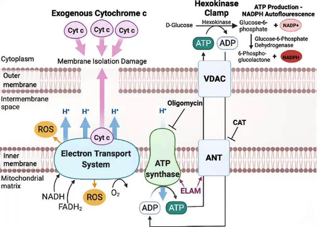
Figure 1 Respiration and membrane potential response to ADP stimulation in isolated muscle mitochondria require supplemental cytochrome c and hexokinase clamp. ADP/ATP transport pathway and binding of ELAM to ATP Synthase and ANT.
I. Overview of the Relationship Between Mitochondria and Aging
Mitochondria, as the cell’s powerhouse, are responsible for synthesizing the majority of ATP within the cell and are also involved in various metabolic regulation and signal transduction processes. As age increases, mitochondrial function gradually declines, which is one of the key markers of aging. Mitochondrial dysfunction manifests in multiple ways, including the accumulation of mutations in mitochondrial DNA (mtDNA), reduced activity of mitochondrial respiratory chain complexes, leading to insufficient energy production; increased production of reactive oxygen species (ROS) by mitochondria, triggering oxidative stress damage, which further disrupts intracellular biomolecules such as proteins, lipids, and nucleic acids, accelerating cellular aging and death. Abnormal mitochondrial function also disrupts intracellular calcium homeostasis, interfering with normal cellular physiological functions. These changes interact with each other, forming a vicious cycle that collectively drives the aging process.
II. Mechanism of Action of SS-31
Improving Mitochondrial Energy Metabolism
Enhancing ADP Sensitivity: During aging, mitochondrial sensitivity to ADP decreases, impairing ATP synthesis efficiency. Studies indicate that SS-31 can directly bind to the mitochondrial ADP transporter ANT, increasing ANT’s uptake of ADP, thereby enhancing mitochondrial sensitivity to ADP. In aged muscle mitochondria, SS-31 treatment increased ADP uptake via ANT, thereby enhancing mitochondrial respiration under ADP stimulation, increasing ATP production, and improving energy metabolism in aged muscle mitochondria.
Regulation of mitochondrial respiratory chain complexes: Mitochondrial respiratory chain complexes are key components of mitochondrial energy production. During aging, the activity of respiratory chain complexes often decreases. SS-31 may maintain or enhance the activity of respiratory chain complexes by stabilizing their structural integrity or regulating the expression of related proteins. Previous studies have observed that in aging-related mitochondrial dysfunction models, the activity of respiratory chain complexes is restored after SS-31 treatment, suggesting its positive regulatory effect on the mitochondrial respiratory chain.
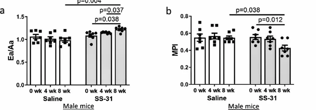
Figure 2 SS-31 treatment reverses cardiac aging phenotypes
Reducing oxidative stress damage
Reducing ROS production: Mitochondria are one of the primary sources of ROS within cells, and ROS production increases in mitochondria during aging. SS-31 can reduce ROS production through multiple pathways. It improves mitochondrial energy metabolism, making the mitochondrial electron transport chain more efficient and reducing electron leakage, thereby lowering ROS production. SS-31 may directly act on the mitochondrial membrane, altering its physical properties, reducing oxidative damage sites on the membrane, and inhibiting ROS production. In cardiac cells from aged mice, treatment with SS-31 significantly reduced mitochondrial ROS levels, indicating its efficacy in reducing ROS production.
Regulation of the antioxidant enzyme system: Cells contain an antioxidant enzyme system, including superoxide dismutase (SOD), catalase (CAT), and glutathione peroxidase (GSH-Px), which are responsible for scavenging ROS. Research suggests that SS-31 may enhance cellular antioxidant defense capabilities by upregulating the expression or activity of these antioxidant enzymes. In certain cellular models, treatment with SS-31 resulted in a significant increase in the activity of SOD and GSH-Px, aiding in the timely clearance of excess ROS within cells and alleviating oxidative stress-induced damage.
Maintaining mitochondrial membrane stability
Binding to mitochondrial membrane phospholipids: SS-31 has a unique structure that allows it to preferentially bind to regions of the mitochondrial membrane rich in phosphatidylserine. Phosphatidylserine is a phospholipid specific to the mitochondrial membrane and is crucial for maintaining the structural and functional integrity of the mitochondrial membrane. During aging, phosphatidylserine is susceptible to oxidative damage, leading to abnormalities in mitochondrial membrane structure and function. After binding to phosphatidylserine, SS-31 may protect phosphatidylserine from oxidation while stabilizing mitochondrial membrane structure, preventing a decrease in mitochondrial membrane potential and the opening of the mitochondrial permeability transition pore (mPTP). In vitro experiments have shown that SS-31 effectively inhibits oxidative stress-induced decreases in mitochondrial membrane potential and mPTP opening, thereby maintaining mitochondrial membrane stability.
Regulation of membrane-associated proteins: The mitochondrial membrane contains various proteins involved in mitochondrial material transport, energy metabolism, and signal transduction processes. SS-31 may indirectly influence mitochondrial membrane stability by regulating the activity or expression of these membrane-associated proteins. It can regulate the function of voltage-dependent anion channels (VDAC), which are important channel proteins on the outer mitochondrial membrane and play a key role in maintaining material exchange and signal transmission between mitochondria and the cytoplasm. SS-31’s regulation of VDAC helps maintain normal mitochondrial membrane function and stability.
III. Specific effects of SS-31 in anti-aging
Effects on the cardiovascular system
Improving cardiac function: The heart is a high-energy-demand organ, and mitochondrial function is critical for cardiac function. In aged mice, the heart often exhibits age-related issues such as diastolic dysfunction. Scientists have found that after 8 weeks of SS-31 treatment, the diastolic function of aged mice’s hearts was significantly improved. This improvement was accompanied by the normalization of mitochondrial proton leakage in myocardial cells, reduced mitochondrial ROS levels, decreased cardiac protein oxidation levels, and a shift in the thiol redox state of proteins toward a more reduced state. SS-31 also increased the phosphorylation level of cMyBP-C Ser282 in myocardial cells, which is closely associated with improved cardiac diastolic function.
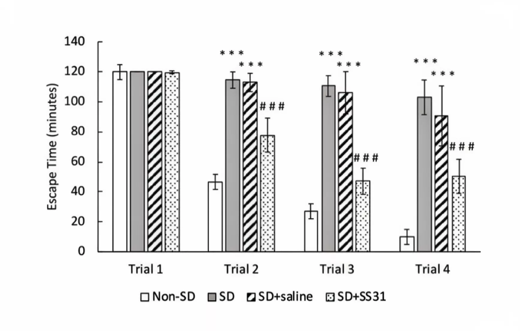
Figure 3 Learning impairment in aging mice induced by sleep deprivation was prevented with SS31.
Vascular protection: During aging, endothelial cell function is impaired, vascular elasticity decreases, and cardiovascular diseases are more likely to occur. In a hypertension-induced microbleeding model in the brains of aged mice, treatment with SS-31 exhibits significant vascular protective effects. It reduces mitochondrial free radical production induced by hypertension, alleviates oxidative stress damage to the vascular wall, thereby significantly delaying the onset of microbleeding and reducing its incidence. Therefore, SS-31 plays an important role in maintaining the normal structure and function of blood vessels and preventing age-related vascular diseases.
Effects on the nervous system
Alleviating cognitive dysfunction: As age increases, nervous system function gradually declines, and cognitive dysfunction such as memory impairment and reduced learning ability becomes increasingly prominent. Mitochondrial dysfunction plays a crucial role in the onset and progression of neurodegenerative diseases and cognitive dysfunction. In a model of cognitive dysfunction induced by isoflurane in aged mice, SS-31 can reverse mitochondrial dysfunction, rescue isoflurane-induced cognitive deficits. SS-31 promotes the regulation of BDNF signaling, reverses the downregulation of synaptic plasticity-related proteins such as synaptophysin, PSD-95, and p-CREB, and upregulates NR2A, NR2B, CaMKIIα, and CaMKIIβ, thereby enhancing synaptic plasticity and protecting cognitive function.
Alleviating the negative effects of sleep deprivation: Sleep deprivation is a common stressor, and its negative effects on the nervous system become more pronounced with age, leading to cognitive dysfunction and increased risk of neurodegenerative diseases. In 20-month-old aged mice, SS-31 (3 mg/kg) was administered via subcutaneous injection daily for 4 consecutive days, with 4 hours of sleep deprivation on the last two days. Results showed that sleep-deprived mice treated with SS-31 exhibited no significant impairment in learning ability, with restored brain mitochondrial ATP levels and synaptic plasticity-regulating proteins, as well as reduced levels of reactive oxygen species (ROS) and inflammatory cytokines in the hippocampus. This suggests that SS-31 may have potential therapeutic benefits in mitigating the adverse neurological effects of short-term sleep deprivation in aged mice.
Effects on the renal system: In the kidneys of aged mice, glomerulosclerosis is associated with mitochondrial damage in glomerular epithelial cells. After 8 weeks of SS-31 treatment in 26-month-old aged mice, SS-31 improved age-related mitochondrial morphology and alleviated glomerulosclerosis. Specifically, it reduced the expression of aging markers (p16, aging-associated β-Gal), increased the density of apical epithelial cells, and decreased the expression of markers of parietal epithelial cell activation (collagen IV, pERK1/2, and α-smooth muscle actin). Although SS-31 did not affect podocyte density, it reduced markers of podocyte damage (desmin), improved cytoskeletal integrity (synaptophysin), and was accompanied by higher glomerular endothelial cell density (CD31). This suggests that short-term SS-31 treatment also has a protective effect on glomerular mitochondria and improves glomerular structure.
Effects at the cellular level
Delaying cellular aging: In cell experiments, H₂O₂ was used to induce a stress-induced aging model in HEK293T cells, followed by intervention with SS-31. The results showed that the SA-β-gal positive rate was significantly reduced in the SS-31 group, indicating a decrease in cellular aging levels. Additionally, the intracellular ROS fluorescence intensity decreased, mitochondrial membrane potential increased, and ATP levels rose in the SS-31 group. Protein immunoblotting analysis revealed that the expression levels of P53, P21, and Acetyl-p53 proteins were higher in the model group compared to the control group, while the SS-31 group showed a decrease compared to the model group. Conversely, the expression level of Sirt1 protein was lower in the model group compared to the control group, while the SS-31 group showed an increase compared to the model group. Thus, it can be concluded that SS-31 can delay HEK293T cell aging by improving mitochondrial function and regulating the expression of aging-related proteins within cells.
Protection against oxidative stress-induced cellular damage: In the H₂O₂-induced oxidative stress damage model of ARPE-19 cells, treatment with SS-31 significantly increased cell survival rate, reduced intracellular ROS levels, decreased the proportion of cells with reduced mitochondrial membrane potential, markedly lowered PI positivity rate (reflecting the extent of cell death), and significantly attenuated the upregulation of RIP3 protein expression. These results indicate that SS-31 has a significant protective effect against H₂O₂-induced oxidative stress damage in ARPE-19 cells, which may aid in counteracting age-related damage.
Conclusion
The mitochondrial-targeted peptide SS-31 contributes to anti-aging. From its multifaceted regulatory mechanisms on mitochondrial function to its significant anti-aging effects at various systemic and cellular levels, it demonstrates potential as an important anti-aging strategy.
Sources
[1] Patai R, Patel K, Csik B, et al. Aging, mitochondrial dysfunction, and cerebral microhemorrhages: a preclinical evaluation of SS-31 (elamipretide) and development of a high-throughput machine learning-driven imaging pipeline for cerebromicrovascular protection therapeutic screening[J]. Geroscience, 2025.DOI:10.1007/s11357-025-01634-5.
[2] Pharaoh G, Kamat V, Kannan S, et al. The mitochondrially targeted peptide elamipretide (SS-31) improves ADP sensitivity in aged mitochondria by increasing uptake through the adenine nucleotide translocator (ANT)[J]. Geroscience, 2023,45(6):3529-3548.DOI:10.1007/s11357-023-00861-y.
[3] Chiao Y A, Zhang H, Sweetwyne M, et al. Late-life restoration of mitochondrial function reverses cardiac dysfunction in old mice[J]. Elife, 2020,9.DOI:10.7554/eLife.55513.
[1] Wu J, Dou Y, Ladiges W C. Adverse Neurological Effects of Short-Term Sleep Deprivation in Aging Mice Are Prevented by SS31 Peptide[J]. Clocks & Sleep, 2020,2(3):325-333.DOI:10.3390/clockssleep2030024.
[4] Sweetwyne M T, Pippin J W, Eng D G, et al. The mitochondrial-targeted peptide, SS-31, improves glomerular architecture in mice of advanced age[J]. Kidney International, 2017,91(5):1126-1145.DOI:10.1016/j.kint.2016.10.036.
[5] Wu J, Zhang M, Li H, et al. BDNF pathway is involved in the protective effects of SS-31 on isoflurane-induced cognitive deficits in aging mice[J]. Behavioural Brain Research, 2016,305:115-121.DOI:10.1016/j.bbr.2016.02.036.
Product available for research use only:
<
By Peplyte 1 month ago
ALL ARTICLES AND PRODUCT INFORMATION PROVIDED ON THIS WEBSITE ARE SOLELY FOR INFORMATION DISSEMINATION AND EDUCATIONAL PURPOSES.
The products provided on this website are intended exclusively for in vitro research. In vitro research (Latin: *in glass*, meaning in glassware) is conducted outside the human body. These products are not pharmaceuticals, have not been approved by the U.S. Food and Drug Administration (FDA), and must not be used to prevent, treat, or cure any medical condition, disease, or ailment. It is strictly prohibited by law to introduce these products into the human or animal body in any form.
Aging, as an inevitable natural process in living organisms, has long been a focal point of scientific research. As organisms age, cells gradually enter a senescent state, and the accumulation of these senescent cells can negatively impact the normal functioning of tissues and organs, leading to various age-related diseases. FOX04 – DRI is beneficial for clearing senescent cells.
Overview of FOX04 – DRI
FOX04 – DRI is a synthetic peptide, with the full name fork head box O transcription factor 4 – D – Retro – Inverso. Its discovery stems from in-depth research into the regulatory mechanisms of senescent cells. During cellular senescence, multiple molecular mechanisms are involved, and the FOX04 transcription factor plays a key role in this process. FOX04 exhibits specific expression patterns and functional changes in senescent cells. By elucidating its mechanism of action, scientists designed and synthesized FOX04-DRI to specifically target senescent cells, aiming to address age-related issues.
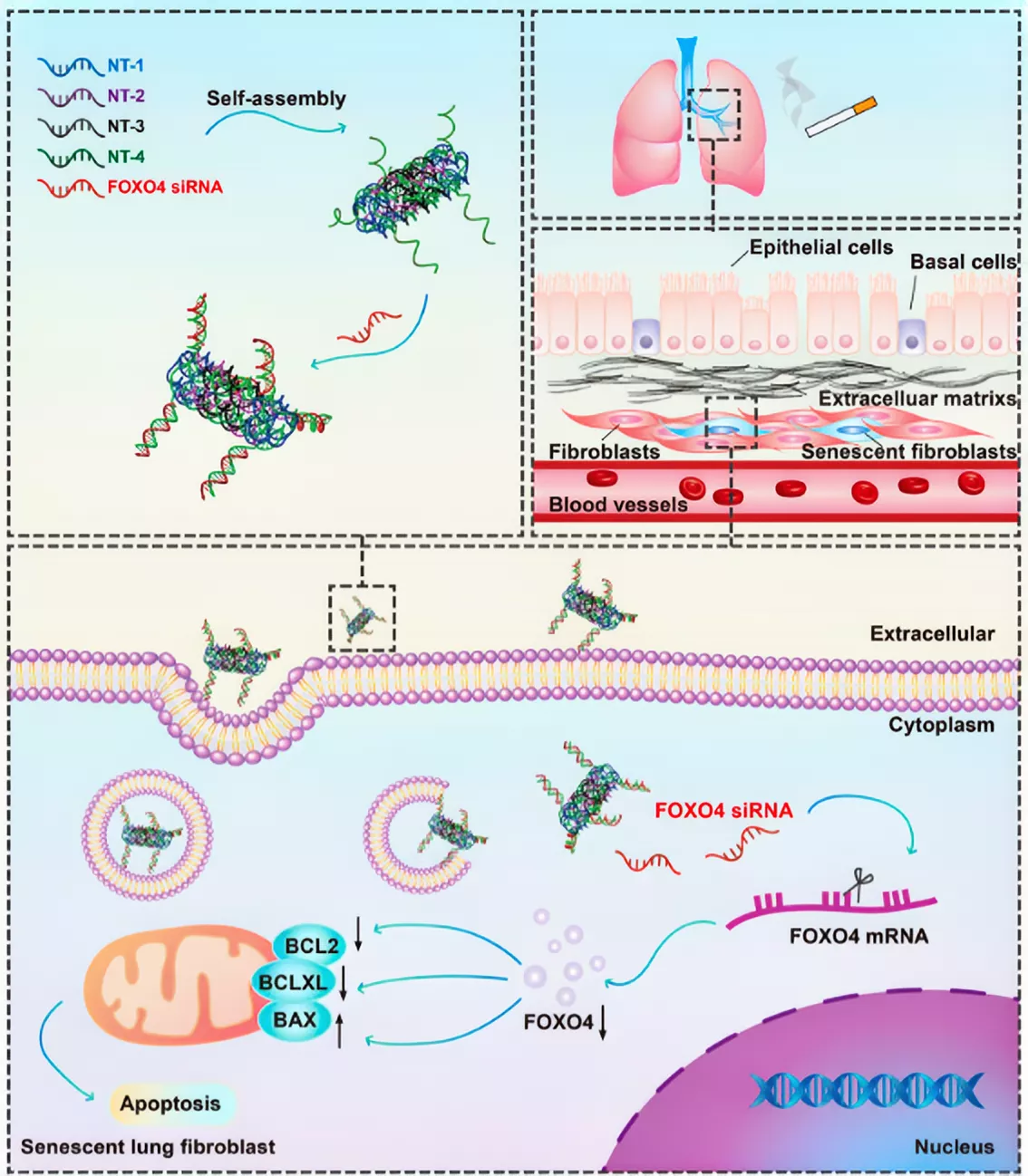
Figure 1 Schematic illustration of the elimination of cigarette smoke-induced senescent lung fibroblasts using FOXO4 siRNA-loaded DNA nanoparticles.
Mechanism of FOX04-DRI in clearing senescent cells
Interference with FOX04-p53 interaction: In senescent cells, FOX04 interacts with the p53 protein. The p53 protein plays a central role in cellular stress responses, cell cycle regulation, and apoptosis. Under normal conditions, p53 promotes apoptosis in damaged or senescent cells by activating the expression of a series of apoptosis-related genes, thereby maintaining the health of the cell population. In senescent cells, FOX04 binds to p53, inhibiting its transport to the cell nucleus, preventing p53 from performing its pro-apoptotic function, resulting in the survival and accumulation of senescent cells. FOX04-DRI can specifically interfere with this interaction between FOX04 and p53, blocking their binding, allowing p53 to re-enter the cell nucleus, activate the apoptosis signaling pathway, and induce apoptosis in senescent cells, thereby achieving the clearance of senescent cells.
Regulation of intracellular signaling pathways: In addition to interfering with the FOX04-p53 interaction, FOX04-DRI may also influence the fate of senescent cells by regulating other intracellular signaling pathways. The PI3K/AKT signaling pathway plays a key regulatory role in various aspects of cell growth, proliferation, survival, and metabolism. Studies have shown that the activity of the PI3K/AKT signaling pathway is often altered in senescent cells, affecting their normal functions. FOX04-DRI may regulate the activity of the PI3K/AKT signaling pathway, altering the balance of signal transduction within senescent cells, thereby creating an unfavorable intracellular environment for their survival and inducing apoptosis. Regulation of this signaling pathway may also influence cellular metabolic states, further driving senescent cells toward apoptosis.
Influence on the Senescence-Associated Secretory Phenotype (SASP): Senescent cells secrete a series of cytokines, chemokines, and proteases, forming the senescence-associated secretory phenotype (SASP). SASP not only negatively affects surrounding normal cells, disrupting their normal functions, but also triggers chronic inflammatory responses, further accelerating the aging process of tissues and organs. FOX04-DRI may mitigate the adverse effects of senescent cells on the surrounding microenvironment by inhibiting the expression and secretion of SASP-related factors in senescent cells. In the study, it was found that after treating senescent cells with FOX04-DRI, the expression levels of SASP-related factors such as IL-1α, IL-1β, tumor necrosis factor α (TNFα), and MMP2 in senescent cells were significantly reduced. This indicates that FOX04-DRI can effectively regulate the SASP of senescent cells, improve the cellular microenvironment, and indirectly promote the clearance of senescent cells and the recovery of the tissue microenvironment.
The Role of FOX04-DRI in Different Tissues and Diseases
Role in the Reproductive System: Male late-onset hypogonadism is an age-related disease whose core mechanism is dysfunction of aged testicular interstitial cells. FOX04 is specifically expressed in human testicular interstitial cells, and in the elderly, the nuclear translocation of FOX04 is associated with reduced testosterone synthesis. Using hydrogen peroxide-induced aged TM3 testicular interstitial cells as an in vitro model, it was observed that FOX04 can maintain the viability of aged testicular interstitial cells and inhibit their apoptosis. FOX04-DRI selectively induces p53 nuclear export and apoptosis of aged testicular interstitial cells by disrupting the FOX04-p53 interaction. In a natural aging mouse model, administration of FOX04-DRI improved the testicular microenvironment and alleviated age-related testosterone secretion deficiency. Therefore, FOX04-DRI may be beneficial for the treatment of male late-onset hypogonadism.
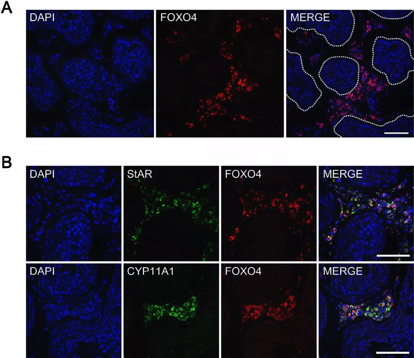
Figure 2 Expression of FOXO4 in human testes detected by immunofluorescent staining.
Role in the respiratory system
Pulmonary fibrosis: Pulmonary fibrosis (PF) is a progressive interstitial lung disease. Its incidence and prevalence increase with age, and cellular senescence is considered one of the key driving factors in its pathogenesis. In a bleomycin (BLM)-induced PF mouse model, FOX04-DRI demonstrated effects similar to those of the approved drug pirfenidone. It reduces the number of senescent cells, downregulates the expression of the senescence-associated secretory phenotype (SASP), and alleviates BLM-induced morphological changes and collagen deposition in lung tissue. Additionally, FOX04-DRI increases the proportion of type 2 alveolar epithelial cells (AEC2) and fibroblasts while reducing the number of myofibroblasts. In vitro experiments showed that compared to mouse and human lung fibroblast cell lines, FOX04-DRI was more inclined to kill transforming growth factor-β (TGF-β)-induced myofibroblasts, and by inhibiting myofibroblasts, it led to the downregulation of extracellular matrix (ECM) receptor interaction pathways in BLM-induced PF.
Chronic obstructive pulmonary disease (COPD): The pathogenesis and progression of COPD are closely associated with cellular senescence. In cigarette smoke-induced senescent pulmonary fibroblasts, FOX04 expression is elevated. To knockdown FOX04 in senescent fibroblasts, self-assembling DNA nanotubes (siFOXO4-NT) loaded with single-stranded FOX04 siRNA were designed and synthesized. Studies have shown that siFOXO4-NT can enter human lung fibroblasts (HFL-1 cells) in a concentration- and time-dependent manner, thereby reducing FOX04 levels in vitro. siFOXO4-NT selectively eliminates aged HFL-1 cells by reducing the elevated BCLXL expression and BCL2/BAX ratio in cigarette smoke extract (CSE)-induced aged HFL-1 cells.
Role in cartilage tissue: Autologous chondrocyte implantation (ACI) is a method for treating joint cartilage damage and preventing post-traumatic osteoarthritis. During the in vitro expansion of chondrocytes, a necessary step in ACI, senescent cells are generated, which negatively impact the quality and quantity of newly formed cartilage. Researchers hypothesized that FOX04-DRI treatment could remove senescent cells from expanded chondrocytes, thereby enhancing their potential to generate high-quality cartilage. To simulate the in vitro expansion process in ACI, chondrocytes isolated from healthy donors were expanded to population doubling level (PDL) 9, representing chondrocytes ready for implantation, while cells at PDL3 served as a minimal expansion control. Results showed that FOX04-DRI treatment removed over half of the cells at PDL9 but had no significant effect on the number of PDL3 chondrocytes. The senescence level of PDL9 chondrocytes treated with FOX04-DRI was significantly reduced. Based on standard micro-cluster culture results, FOX04-DRI pretreatment did not enhance the chondrogenic potential of PDL9 chondrocytes, but chondroid tissue derived from FOX04-DRI-pretreated PDL9 cells exhibited lower expression of aging-associated secretory factors. FOX04-DRI effectively removed senescent cells from PDL9 chondrocytes.
Role in radiation-induced pulmonary fibrosis (RIPF): RIPF is a common and severe complication following thoracic tumor radiotherapy. C57BL/6 mice were randomly divided into irradiation and irradiation + FOX04-DRI groups, with the right hemithorax of each group receiving 17 Gy X-ray irradiation. FOX04-DRI group and irradiation + FOX04-DRI group mice were administered FOX04-DRI via intraperitoneal injection at weeks 16 and 20 post-irradiation. Results showed that FOX04-DRI reduced collagen deposition in lung tissue of RIPF mice, decreased expression of col1α1 and α-SMA, and reduced the number of β-galactosidase (β-gal) positive cells (a marker of senescent cells) in lung tissue. FOX04-DRI was able to inhibit the expression of P21 and P16^(Ink4a) genes and proteins in lung tissue of RIPF mice, as well as the expression of SASP genes IL-1α, IL-1β, tumor necrosis factor α (TNFα), and MMP2. FOX04-DRI also reduces reactive oxygen species (ROS) levels in lung tissue of RIPF mice and promotes the activation of p-AKT and p-PI3K proteins. This suggests that FOX04-DRI reduces oxidative stress and inhibits cellular senescence by activating the PI3K/AKT signaling pathway, thereby reversing RIPF.
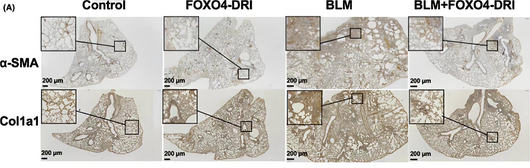
Figure 3 FOXO4-DRI alleviates myofibroblast differentiation in BLM-induced PF mice.
Conclusion
In summary, FOX04-DRI, as a revolutionary drug capable of specifically eliminating senescent cells, plays an important role in the treatment of age-related diseases and aging intervention.
Sources
[1] Han Y, Wu Y, He B, et al. DNA nanoparticles targeting FOXO4 selectively eliminate cigarette smoke-induced senescent lung fibroblasts[J]. Nanoscale Advances, 2023,5(21):5965-5973.DOI:10.1039/d3na00547j.
[2] Han X, Yuan T, Zhang J, et al. FOXO4 peptide targets myofibroblast ameliorates bleomycin-induced pulmonary fibrosis in mice through ECM-receptor interaction pathway[J]. Journal of Cellular and Molecular Medicine, 2022,26(11):3269-3280.DOI:10.1111/jcmm.17333.
[3] Huang Y, He Y, Makarcyzk M J, et al. Senolytic Peptide FOXO4-DRI Selectively Removes Senescent Cells From in vitro Expanded Human Chondrocytes[J]. Frontiers in Bioengineering and Biotechnology, 2021,9:677576.DOI:10.3389/fbioe.2021.677576.
[4] Zhang C, Xie Y, Chen H, et al. FOXO4-DRI alleviates age-related testosterone secretion insufficiency by targeting senescent Leydig cells in aged mice[J]. Aging (Albany Ny), 2020,12(2):1272-1284.DOI:10.18632/aging.102682.
[5] Baar M P. DNA Damage Induced Cell Fates during Aging, 2019[C]. https://api.semanticscholar.org/CorpusID:196682180
Product available for research use only:

<
By Peplyte 18 days ago
ALL ARTICLES AND PRODUCT INFORMATION PROVIDED ON THIS WEBSITE ARE SOLELY FOR INFORMATION DISSEMINATION AND EDUCATIONAL PURPOSES.
The products provided on this website are intended exclusively for in vitro research. In vitro research (Latin: *in glass*, meaning in glassware) is conducted outside the human body. These products are not pharmaceuticals, have not been approved by the U.S. Food and Drug Administration (FDA), and must not be used to prevent, treat, or cure any medical condition, disease, or ailment. It is strictly prohibited by law to introduce these products into the human or animal body in any form.
Overview of Triple Agonists
In recent years, with the deepening of research into the pathogenesis of metabolic diseases, the development of multi-target agonist drugs targeting multiple hormone receptors has become a hot topic. Retatrutid is a novel triple agonist of the glucose-dependent insulinotropic polypeptide (GIP), glucagon-like peptide-1 (GLP-1), and glucagon (GC) receptors.
(1) Physiological Basis of Hormone Receptor Functions
GIP Receptor: GIP is an intestinal insulin-secreting hormone secreted by K cells in the small intestine, released rapidly after meals. Upon binding to the GIP receptor, GIP stimulates insulin secretion, and this stimulatory effect is glucose-dependent, meaning that as blood glucose levels rise, GIP enhances its insulin-secreting effect, helping to maintain stable blood glucose levels. Additionally, GIP participates in the regulation of lipid metabolism, influencing adipocyte differentiation and lipid accumulation.
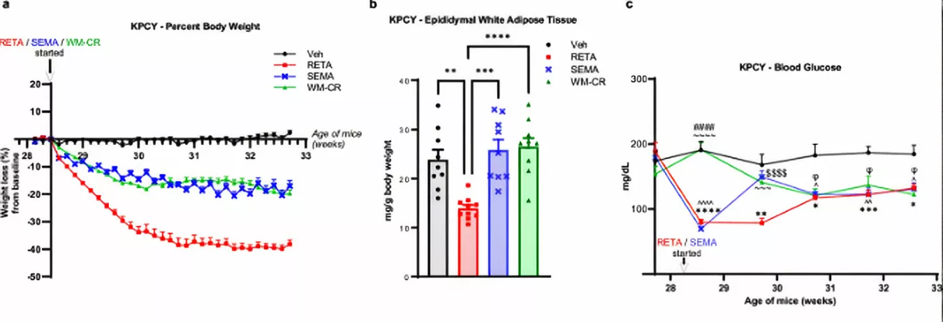
Fig. 1. RETA or SEMA treatment reduces body weight and improves fasting blood glucose.a Percent changes in body weight from baseline are reported. b, Epididymal white adipose tissue weight was quantified at the endpoint. c Fasting blood glucose was quantified at baseline and throughout the study at indicated time points until the endpoint. d Tumor engraftment or “tumor take” is reported.
GLP-1 receptor: GLP-1 is secreted by intestinal L cells and also has a glucose-dependent insulinotropic effect, enhancing insulin synthesis and release, inhibiting glucagon secretion, and reducing hepatic glucose output. GLP-1 also delays gastric emptying, increases satiety, and thereby reduces food intake. It has protective and proliferative effects on pancreatic beta cells, helping to maintain pancreatic function.
3. GC Receptor:
After binding to the GC receptor, glucagon primarily acts on the liver by promoting glycogenolysis and gluconeogenesis, thereby elevating blood glucose levels. In lipid metabolism, it promotes lipolysis, increasing the release and oxidation of fatty acids for energy. Under normal physiological conditions, glucagon secretion is inhibited when blood glucose levels rise to maintain glucose balance.
(2) Advantages of Triple Agonist Action
As a triple agonist, Retatrutid can simultaneously activate these three receptors, exerting synergistic effects. By activating GIP and GLP-1 receptors, it enhances glucose-dependent insulin secretion, more effectively lowering blood glucose levels. The increased satiety and delayed gastric emptying induced by GLP-1 receptor activation, along with the lipolysis promoted by GC receptor activation, collectively contribute to weight management. This multi-targeted mechanism of action provides a more comprehensive regulation of various pathophysiological processes associated with metabolic diseases.
Mechanism of Action of Retatrutid in Metabolic Diseases
Metabolic diseases are a class of conditions caused by disruptions in metabolic processes within the body, including common conditions such as diabetes, obesity, and non-alcoholic fatty liver disease. Retatrutid exerts its effects on these metabolic diseases through its unique triple agonist properties.
(1) Mechanism of Action in Diabetes
Glucose Regulation: Retatrutid activates GIP and GLP-1 receptors, enhancing glucose-dependent insulin secretion. When blood glucose levels rise, it more effectively stimulates pancreatic beta cells to secrete insulin, promoting glucose uptake and utilization, thereby lowering blood glucose levels. By inhibiting glucagon secretion and reducing hepatic glucose output, it further stabilizes blood glucose levels. In clinical trials involving patients with type 2 diabetes, Retatrutid treatment resulted in significant reductions in both fasting and postprandial blood glucose levels.
Protection of pancreatic beta cells: Activation of the GLP-1 receptor not only promotes insulin secretion but also has a protective and proliferative effect on pancreatic beta cells. Retatrutid may achieve this by continuously activating the GLP-1 receptor, slowing down beta cell apoptosis, increasing their number and function, thereby improving insulin secretion capacity and maintaining stable blood glucose levels over the long term.
(2) Mechanism of action in obesity
Regulation of energy intake: Retatrutid activates GLP-1 receptors, delaying gastric emptying, prolonging the retention time of food in the stomach, increasing satiety, and reducing food intake. It can act on the central nervous system to regulate appetite-related neurotransmitters, such as inhibiting the release of orexin, further reducing appetite, and thereby controlling energy intake.
Increased Energy Expenditure: Activation of the GC receptor promotes lipolysis, increasing the release and oxidation of fatty acids for energy production. The oxidation of fatty acids provides energy to the body, reduces fat storage, and aids in weight loss. Activation of the GIP receptor also participates in energy metabolism regulation, increasing energy expenditure.
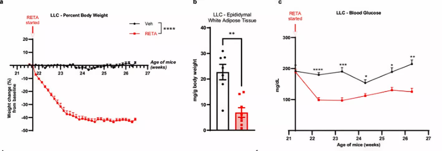 Figure 2 RETA treatment reduces body weight and adiposity, improves fasting blood glucose. a Percent changes in body weight from baseline are reported. b Epididymal white adipose tissue was weighed at the endpoint and normalized to total body weight. c Fasting blood glucose was quantified at baseline and throughout the study at indicated time points until the endpoint.
(3) Mechanism of action in non-alcoholic fatty liver disease
Improved hepatic lipid metabolism: Retatrutid activates the GC receptor, promoting the breakdown and β-oxidation of fat in the liver, thereby reducing fat accumulation in the liver. Activation of the GIP and GLP-1 receptors may regulate the expression of genes related to lipid metabolism in the liver, upregulating the expression of fatty acid transporters and fatty acid oxidases, thereby promoting fatty acid uptake and oxidation, and further improving hepatic lipid metabolism.
Enhanced insulin sensitivity: While improving glucose regulation and increasing insulin secretion, Retatrutid can enhance hepatic insulin sensitivity by activating GLP-1 receptors. Enhanced insulin sensitivity enhances the liver’s response to insulin, better inhibiting hepatic glucose output, reducing de novo fatty acid synthesis, and thereby alleviating hepatic steatosis.
Applications and Effects of Retatrutid in Metabolic Diseases
(1) Applications in Diabetes
lycemic Control Effects: Retatrutid demonstrates significant glycemic control effects in patients with type 2 diabetes. According to data, patients treated with Retatrutid exhibited a significant reduction in hemoglobin A1c (HbA1c) levels. In a randomized controlled trial involving 353 patients with type 2 diabetes, the HbA1c levels in the Retatrutid treatment group decreased by 1.64% from baseline, while there was no significant change in the placebo group.
Comparison with Other Drugs: Compared with traditional antidiabetic drugs such as metformin or GLP-1 receptor agonists, Retatrutid demonstrates superior blood glucose control effects.
(2) Application in obesity
Weight loss effects: Retatrutid demonstrates significant efficacy in the treatment of obesity. In clinical trials targeting adult obese patients, significant weight loss was observed after 48 weeks of treatment with different doses of Retatrutid. In one trial, patients treated with a 12mg dose of Retatrutid experienced an average weight loss of 24.2% after 48 weeks, compared to only 2.1% in the placebo group. Additionally, 83% of participants in the high-dose group achieved weight loss of 15% or more, far exceeding the 2% rate in the placebo group.
Long-Term Effects: Retatrutid maintains weight loss effects well throughout the treatment period. This suggests that long-term use may help maintain stable weight and prevent weight regain, which is beneficial for the long-term management of obesity.
(3) Application in non-alcoholic fatty liver disease
Liver fat reduction: In patients with metabolic dysfunction-associated fatty liver disease and liver fat content ≥10%, Retatrutid treatment resulted in a significant reduction in liver fat content. In a randomized, double-blind, placebo-controlled trial, at 24 weeks, liver fat content decreased by 81.4% and 82.4% from baseline in the 8 mg and 12 mg dose groups, respectively, while the placebo group increased by only 0.3%.
Advantages and Potential of the Triple Agonist
Retatrutid comprehensively regulates multiple pathophysiological pathways of metabolic diseases by simultaneously activating GIP, GLP-1, and GC receptors. From blood glucose control, weight management, to improved hepatic lipid metabolism, this multi-targeted mechanism of action theoretically offers a more comprehensive correction of metabolic disorders compared to single-target drugs, demonstrating potential to address the complex etiology of metabolic diseases. Existing clinical trial data show that Retatrutid has achieved significant effects in the treatment of metabolic diseases such as diabetes, obesity, and non-alcoholic fatty liver disease.
Conclusion
As a novel triple agonist, Retatrutid holds promise for the treatment of metabolic diseases.
Sources
[1] Allard C, Cota D, Quarta C. Poly-Agonist Pharmacotherapies for Metabolic Diseases: Hopes and New Challenges[J]. Drugs, 2024,84(2):127-148.DOI:10.1007/s40265-023-01982-6.
[2] Kaur M, Misra S. A review of an investigational drug retatrutide, a novel triple agonist agent for the treatment of obesity[J]. European Journal of Clinical Pharmacology, 2024,80(5):669-676.DOI:10.1007/s00228-024-03646-0.
[3] Lopez D C, Pajimna J T, Milan M D, et al. 7792 Efficacy of Retatrutid for Weight Reduction and Its Cardiometabolic Effects Among Adults: A Systematic Review and Meta-Analysis[J]. Journal of the Endocrine Society, 2024,8(Supplement_1):bvae163-bvae749.DOI:10.1210/jendso/bvae163.749.
[4] Jastreboff A M, Kaplan L M, Frías J P, et al. Triple-Hormone-Receptor Agonist Retatrutid for Obesity – A Phase 2 Trial[J]. New England Journal of Medicine, 2023,389(6):514-526.DOI:10.1056/NEJMoa2301972.
[5] Doggrell S A. Is retatrutide (LY3437943), a GLP-1, GIP, and glucagon receptor agonist a step forward in the treatment of diabetes and obesity?[J]. Expert Opinion On Investigational Drugs, 2023,32(5):355-359.DOI:10.1080/13543784.2023.2206560.
Product available for research use only:
<
By Peplyte 15 days ago
ALL ARTICLES AND PRODUCT INFORMATION PROVIDED ON THIS WEBSITE ARE SOLELY FOR INFORMATION DISSEMINATION AND EDUCATIONAL PURPOSES.
The products provided on this website are intended exclusively for in vitro research. In vitro research (Latin: *in glass*, meaning in glassware) is conducted outside the human body. These products are not pharmaceuticals, have not been approved by the U.S. Food and Drug Administration (FDA), and must not be used to prevent, treat, or cure any medical condition, disease, or ailment. It is strictly prohibited by law to introduce these products into the human or animal body in any form.
Overview of BPC-157
BPC-157 is a stable gastrointestinal pentapeptide initially discovered for its anti-ulcer properties. It remains stable in human gastric juice for over 24 hours, enabling it to continuously exert its effects in the gastrointestinal environment and laying the foundation for repairing gastrointestinal and joint damage. Its chemical structur
e is GEPPPGKPADDAGLV, with a molecular weight of 1419. This molecular structure enables it to interact with various biomolecules in the body, thereby exerting important physiological functions.
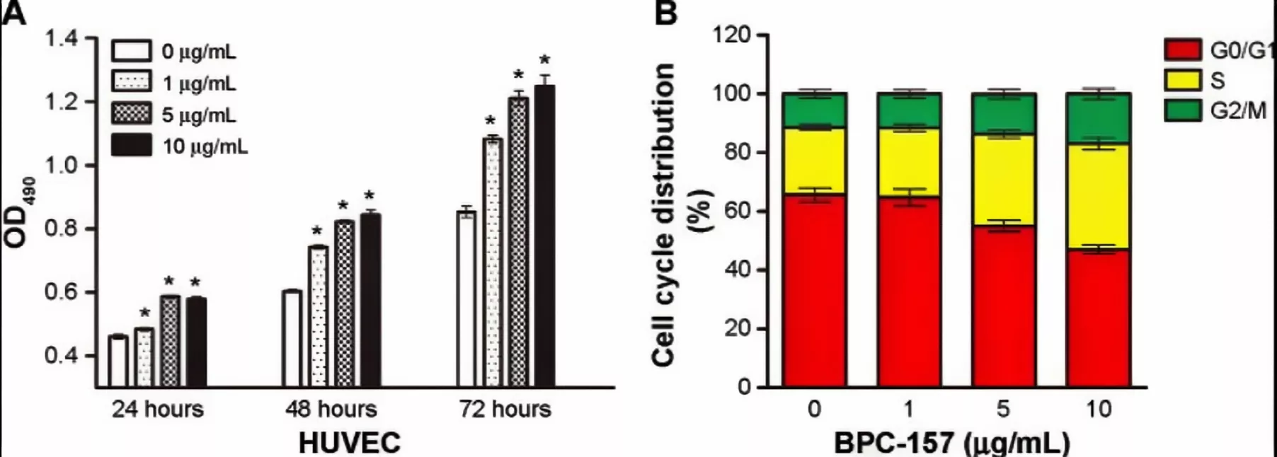
Figure 1 BPC-157 influences the proliferation and cell cycle distribution of HUVECs.
The Repairing Effects of BPC-157 on Gastrointestinal Damage
Protecting Gastric Cells and Maintaining Gastric Integrity
BPC-157 plays a key role in protecting gastric cells, effectively defending against the harmful effects of various drugs on the stomach. Substances such as alcohol and nonsteroidal anti-inflammatory drugs (NSAIDs) often cause damage to gastric cells and disrupt gastric integrity. BPC-157 can protect gastric cells from direct damage by these harmful substances through a series of complex physiological mechanisms. When exposed to alcohol stimulation, BPC-157 regulates metabolic pathways in gastric cells, enhances the cells’ antioxidant defense system, and reduces oxidative damage caused by alcohol metabolites. For NSAID-induced gastric mucosal damage, BPC-157 influences intracellular signaling pathways to inhibit the release of inflammatory mediators, thereby mitigating the destructive effects of inflammatory responses on the gastric mucosa. This helps maintain the integrity of the gastric mucosa and ensure normal physiological functions of the stomach.
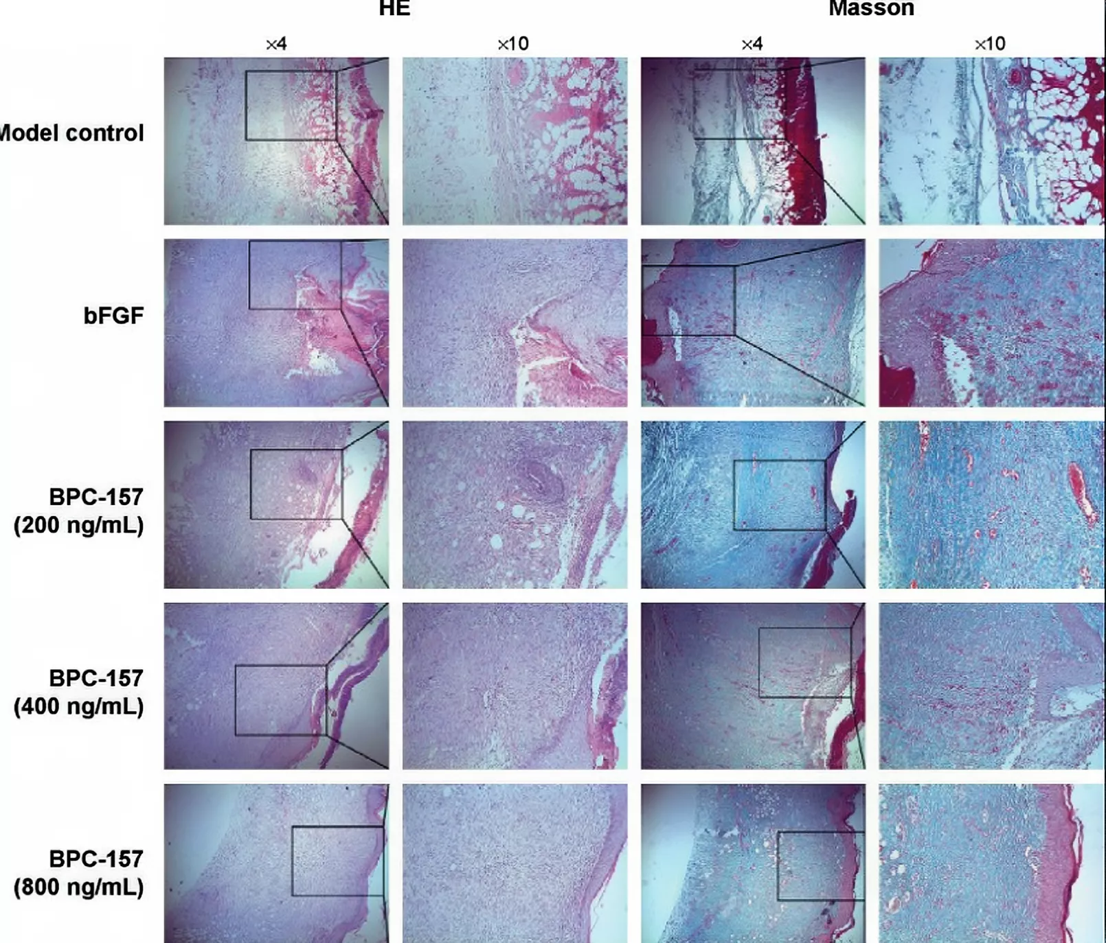
Figure 2 BPC-157 increased wound reepithelialization and collagen content of granulation tissue.
Stabilizing intestinal permeability
Abnormal increases in intestinal permeability are an important factor contributing to the development of many intestinal diseases. Gastrointestinal irritants, such as physical or mental stress, NSAID use, bile acid surfactant stimulation, and alcohol intake, can all lead to increased intestinal epithelial permeability, triggering leaky gut syndrome. BPC-157 can stabilize intestinal permeability by regulating tight junction proteins between intestinal epithelial cells, thereby enhancing intercellular connection strength. According to data, in animal models exposed to the aforementioned stress factors, the use of BPC-157 resulted in increased expression of tight junction proteins between intestinal epithelial cells, with more tightly arranged structures, thereby reducing intestinal permeability to large molecules. This effectively alleviated symptoms of leaky gut syndrome, reduced the risk of harmful substances entering the bloodstream from the intestines, and maintained the stability of the intestinal microenvironment.
Promoting gastrointestinal wound healing
Whether in the healing process of ulcers throughout the gastrointestinal tract or external fistulas (such as esophageal skin fistulas, gastric skin fistulas, duodenal skin fistulas, and colonic skin fistulas) and internal fistulas (such as colonic bladder fistulas and rectal vaginal fistulas) of the gastrointestinal tract, BPC-157 demonstrates significant promotional effects. BPC-157 promotes the proliferation and migration of fibroblasts, accelerates collagen synthesis and deposition, thereby providing the necessary matrix support for wound healing. It also regulates local inflammatory responses, attracts immune cells to the wound site to clear pathogens and necrotic tissue, and promotes angiogenesis, ensuring adequate nutrient and oxygen supply for wound healing. In gastrointestinal anastomosis surgery, when the anastomotic site faces healing difficulties due to factors such as NSAIDs, cystamine, or colectomy, BPC-157 can also play a positive role in promoting anastomotic healing and reducing the risk of complications such as anastomotic fistula.
Effects on pancreatic and gastrointestinal lesions
In a rat model of acute pancreatitis induced by bile duct ligation, BPC-157 demonstrated protective effects on the pancreas whether used as a preventive or therapeutic agent. Prophylactic administration of BPC-157 significantly reduced the severity of pancreatitis, decreased pancreatic necrosis, edema, and neutrophil infiltration, while increasing monocyte counts. In the treatment of severe acute pancreatitis that has already occurred, significant beneficial effects were also observed, with serum amylase levels being controlled. BPC-157 also has a positive impact on concurrent gastric and duodenal lesions, helping to maintain the integrity of the gastrointestinal mucosa and reduce inflammatory damage to the stomach and duodenum.
The Repairing Effects of BPC-157 on Joint Damage
Improvement of temporomandibular joint osteoarthritis
In a rat model of temporomandibular joint osteoarthritis, joint damage was induced surgically, such as unilateral condylar articular surface damage, lateral collateral ligament of the articular disc, and posterior disc attachment level incisions, to simulate the pathological process of osteoarthritis. Following surgery, BPC-157 was administered via intraperitoneal injection. At the 6-month follow-up assessment, the BPC-157 treatment group exhibited a significant increase in overall joint cartilage thickness compared to the control group. Specifically, this manifested as increased thickness in both the cartilage zone and joint zone, reduced cartilage cell aggregation, disappearance of vertical cartilage fissures, and preservation of normal cartilage layering. This suggests that BPC-157 can effectively alleviate the pathological changes of temporomandibular joint osteoarthritis and promote the repair and regeneration of articular cartilage. Its mechanism of action may be related to the interaction of BPC-157 with molecules such as collagen and proteoglycans associated with articular cartilage development. According to research, multiple motifs of BPC-157 are concentrated in collagen α-1 (I), collagen α-1 (IX), aggrecan, fibronectin precursor, chondroitin sulfate proteoglycan NG2 precursor, and chondroitin oligosaccharide precursor, which are crucial for maintaining the structure and function of joint cartilage. BPC-157 may promote the repair and reconstruction of joint cartilage by regulating the expression or activity of these molecules.
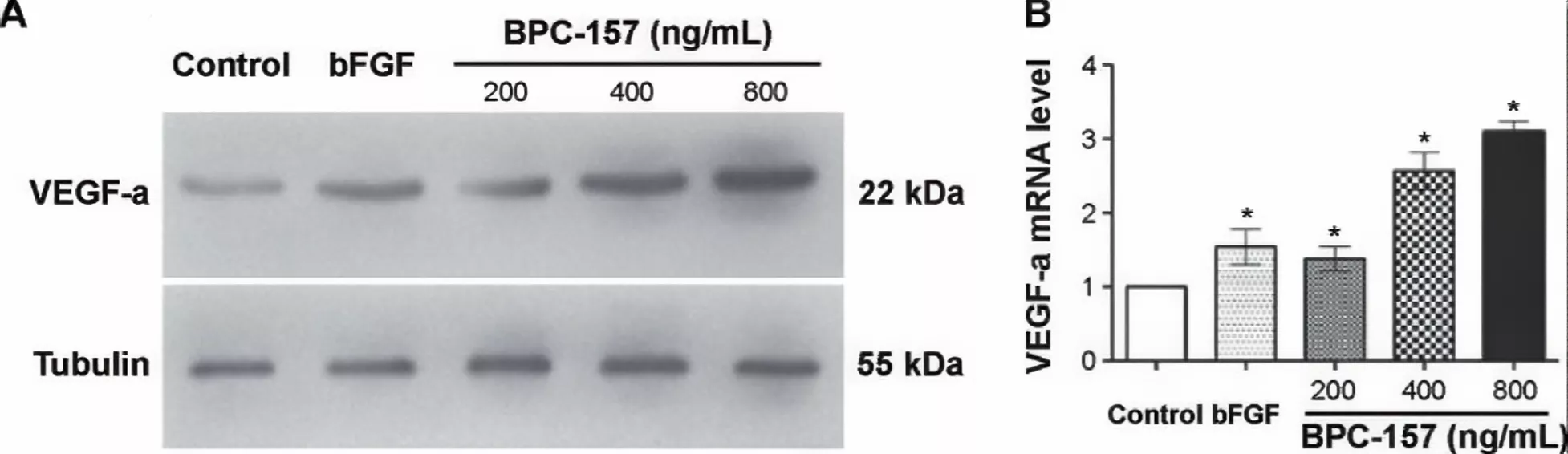
Figure 3 BPC-157 promoted the expression of VEGF-a in wounded skin tissues.
Repair of knee joint injuries
Knee joint pain is a common clinical symptom caused by various factors, such as osteoarthritis, meniscus tears, tendon disorders, ligament tears, or sprains. A retrospective study observed 17 patients with knee joint pain, with 16 patients undergoing follow-up. Among these patients, 12 received intra-articular injections of BPC-157 alone, with 11 (91.6%) experiencing significant improvement in knee pain; the remaining 4 patients received combined injections of BPC-157 and thymosin-β4, with 75% of these patients experiencing significant pain relief. Overall, 87.5% of patients experienced relief of knee pain after using BPC-157 or its combination with thymosin-β4. This indicates that BPC-157 has good efficacy in alleviating various types of knee pain. Further analysis suggests that BPC-157 has the potential to repair tears and promote cartilage generation, which may be an important reason for its efficacy in alleviating knee pain. During the repair process of joint injuries, BPC-157 may promote the proliferation and differentiation of chondrocytes, increase the synthesis of cartilage matrix, thereby repairing damaged cartilage tissue. Additionally, it may also exert a reparative effect on damaged ligaments, tendons, and other tissues, improving joint stability and alleviating pain caused by joint instability.
Repair of tendon-muscle junction injuries
After the tendon of the right quadriceps muscle in rats was separated from the muscle, treatment with BPC-157 demonstrated significant therapeutic effects. From a macroscopic assessment, the control group exhibited a large defect between the quadriceps tendon and muscle that continued to progress, resulting in failed healing. In contrast, rats treated with BPC-157 showed ongoing healing, with the defect being significantly smaller from the outset, ultimately leading to the basic restoration of the tendon-muscle connection. Microscopic examination revealed that the control group exhibited tendon edema, moderate lymphocytic infiltration, and granulation tissue formation by the second week post-surgery, whereas the BPC-157 treatment group showed only mild lymphocytic infiltration, no granulation tissue formation, and good restoration of the tendon-muscle connection structure. Biomechanical assessment showed that muscle strength recovery was poor in the control group, while the BPC-157 treatment group achieved complete recovery. This indicates that BPC-157 not only has therapeutic effects on injured muscles and tendons but also exhibits specific reparative and therapeutic effects on damage to the tendon-muscle junction, aiding in the restoration of normal connection and function between muscles and tendons and ensuring normal joint mobility.
Potential and Prospects of BPC-157 in Clinical Applications
Treatment of Gastrointestinal Diseases
Given BPC-157’s protective effects on gastric cells, stabilization of intestinal permeability, promotion of gastrointestinal wound healing, and positive influence on pancreatic and gastric-duodenal lesions, it holds significant potential in the treatment of various gastrointestinal diseases. In the treatment of gastric ulcers and duodenal ulcers, BPC-157 can accelerate ulcer healing and reduce the risk of recurrence. For inflammatory bowel diseases such as ulcerative colitis, BPC-157 may improve intestinal mucosal barrier function by regulating intestinal immune and inflammatory responses, alleviate symptoms, and promote disease remission. Following gastrointestinal surgery, the use of BPC-157 may help promote anastomotic healing, reduce the incidence of complications, and accelerate the patient’s recovery process.
Treatment of joint diseases
In the field of joint diseases, BPC-157’s reparative effects on temporomandibular joint osteoarthritis and knee injuries provide new insights into its potential application in the treatment of osteoarthritis and sports injuries. For osteoarthritis patients, BPC-157 may serve as a potential disease-modifying drug, not only alleviating pain symptoms but also slowing down cartilage degeneration, promoting cartilage repair, and improving joint function. In the treatment of sports injuries, such as tendon and ligament injuries in the knee and ankle joints commonly seen in athletes, BPC-157 can accelerate the repair of damaged tissues, shorten rehabilitation time, and enhance the recovery speed of athletes’ athletic performance.
Conclusion
As a bioactive peptide with multiple repair functions, BPC-157 demonstrates certain efficacy in the fields of gastrointestinal and joint injury repair.
Sources
[1] Lee E, Padgett B J. Intra-Articular Injection of BPC 157 for Multiple Types of Knee Pain.[J]. Alternative Therapies in Health and Medicine, 2021,27 4:8-13.DOI:https://api.semanticscholar.org/CorpusID:236516759.
[2] Sikiric P, Drmic D, Sever M, et al. Fistulas Healing. Stable Gastric Pentadecapeptide BPC 157 Therapy[J]. Current Pharmaceutical Design, 2020,26(25):2991-3000.DOI:10.2174/1381612826666200424180139.
[3] Sikiric P, Hahm K B, Blagaic A B, et al. Stable Gastric Pentadecapeptide BPC 157, Robert’s Stomach Cytoprotection/Adaptive Cytoprotection/Organoprotection, and Selye’s Stress Coping Response: Progress, Achievements, and the Future[J]. Gut and Liver, 2020,14(2):153-167.DOI:10.5009/gnl18490.
[4] Huang T, Zhang K, Sun L, et al. Body protective compound-157 enhances alkali-burn wound healing in vivo and promotes proliferation, migration, and angiogenesis in vitro[J]. Drug Des Devel Ther, 2015,9:2485-2499.DOI:10.2147/DDDT.S82030.
[5] Kokic N, Sikiric P, Seiwerth S, et al. Pentadecapeptide BPC-157 and rat temporomandibular joint osteoarthrosis[M]. 2001.https://www.researchgate.net/publication/266736896_Pentadecapeptide_BPC-157_and_rat_temporomandibular_joint_osteoarthrosis.
[6] Sikirić P, Seiwerth S, Grabarević Z, et al. Salutary and prophylactic effect of pentadecapeptide BPC 157 on acute pancreatitis and concomitant gastroduodenal lesions in rats[J]. Digestive Diseases and Sciences, 1996,41(7):1518-1526.DOI:10.1007/BF02088582.
Product available for research use only:
By Peplyte 1 month ago
ALL ARTICLES AND PRODUCT INFORMATION PROVIDED ON THIS WEBSITE ARE SOLELY FOR INFORMATION DISSEMINATION AND EDUCATIONAL PURPOSES.
The products provided on this website are intended exclusively for in vitro research. In vitro research (Latin: *in glass*, meaning in glassware) is conducted outside the human body. These products are not pharmaceuticals, have not been approved by the U.S. Food and Drug Administration (FDA), and must not be used to prevent, treat, or cure any medical condition, disease, or ailment. It is strictly prohibited by law to introduce these products into the human or animal body in any form.
Overview
TB 500, also known as Thymosin Beta-4, is a peptide composed of 43 amino acids. It plays a crucial role in various physiological processes in the human body, particularly in muscle recovery. TB 500 was initially isolated from thymus tissue, and research has shown that it has important regulatory functions in cell migration, angiogenesis, and tissue repair.
From a molecular structural perspective, TB 500’s unique amino acid sequence confers specific biological activity. Its structural stability enables it to maintain functionality across diverse physiological environments, laying the foundation for its role in muscle recovery. This peptide is widely distributed throughout the body, with significant amounts present in muscle tissue, indicating a connection to muscle physiological functions.
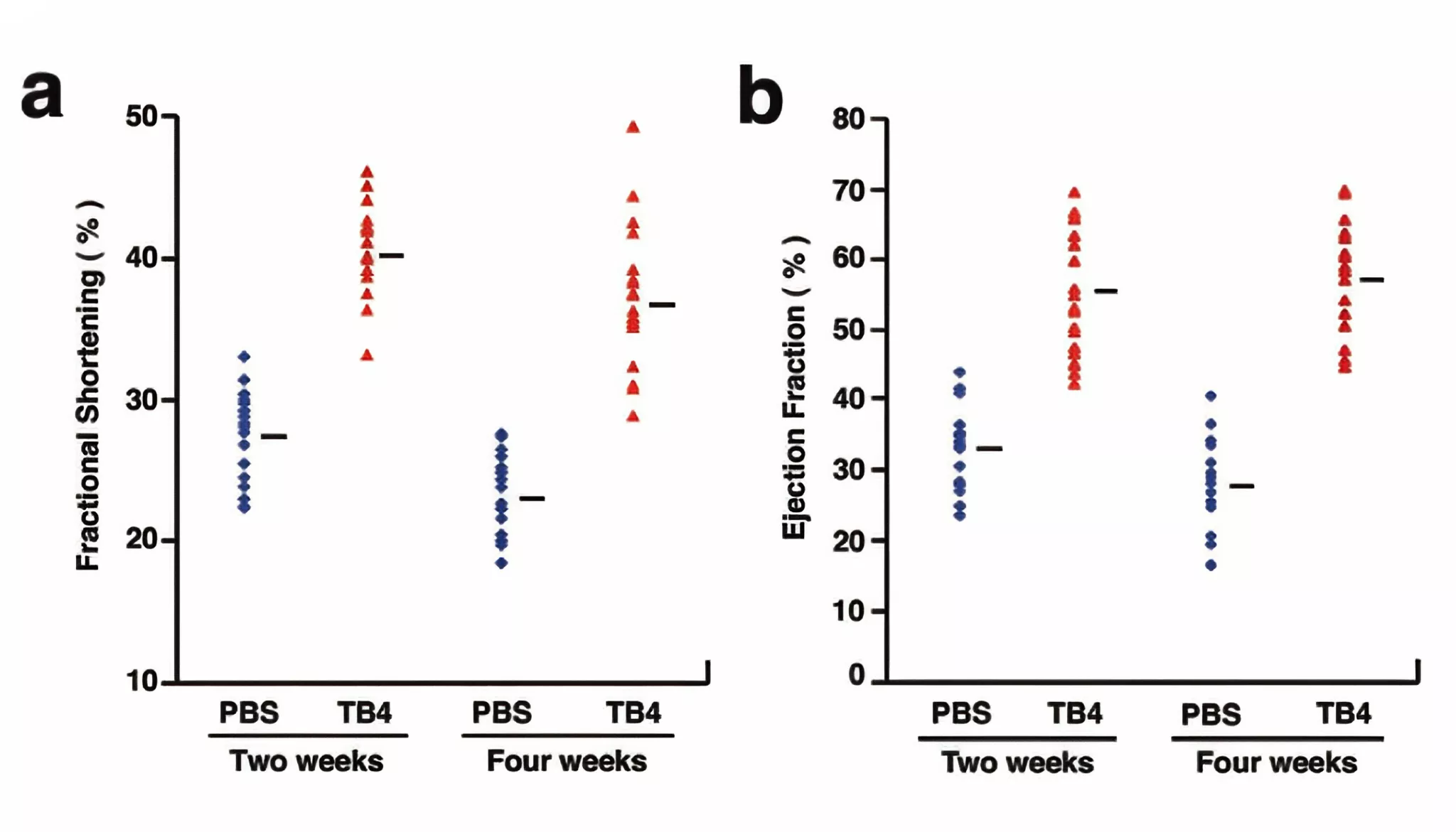
Figure 1 TB4 treatment following coronary ligation improves myocardial function in vivo. Distribution of left ventricular fractional shortening (FS).
Mechanisms by which TB 500 promotes muscle recovery
(1)Cell migration and regeneration mechanisms
Promotion of myoblast migration
Following muscle injury, myoblast migration is critical for repairing the damaged site. TB 500 can bind to specific receptors on the cell membrane, activate intracellular signaling pathways, and promote myoblast migration to the injured site. Studies have shown that in vitro cell experiments, adding TB 500 significantly accelerates myoblast migration speed and increases migration distance. This process involves the remodeling of the cytoskeleton, and TB 500 can regulate the polymerization and depolymerization of actin, enabling myoblasts to move more effectively to the damaged muscle area and provide the necessary cellular source for muscle regeneration.
Satellite Cell Activation and Differentiation
Satellite cells are stem cells in muscle tissue that can be activated and differentiated into myoblasts upon muscle injury, subsequently fusing to form new muscle fibers. TB 500 enhances the activation signals of satellite cells, promoting their transition from a quiescent state to a proliferative state. It regulates the expression of relevant transcription factors, such as MyoD and Myf5, to guide satellite cells toward myoblast differentiation. In animal experiments, local injection of TB 500 into injured muscle resulted in a significant increase in the number of activated satellite cells, as well as improved quantity and quality of newly formed muscle fibers, indicating that TB 500 plays a crucial role in promoting satellite cell-mediated muscle regeneration.
(2)Angiogenesis Mechanism
Induction of Vascular Endothelial Growth Factor (VEGF) Expression
Angiogenesis is crucial for muscle recovery, as it supplies oxygen and nutrients to injured tissues while removing metabolic waste. TB 500 can stimulate muscle cells and surrounding mesenchymal cells to express vascular endothelial growth factor (VEGF). VEGF is a key regulator of angiogenesis, promoting the proliferation, migration, and tubule formation of vascular endothelial cells. Research shows that in muscle tissue treated with TB 500, both mRNA and protein expression levels of VEGF are significantly elevated. This induction is achieved by activating specific intracellular signaling pathways, such as the PI3K-Akt and MAPK signaling pathways. Activation of these pathways promotes the transcription and translation of the VEGF gene, thereby increasing VEGF secretion and facilitating new blood vessel formation, improving blood supply to injured muscle.
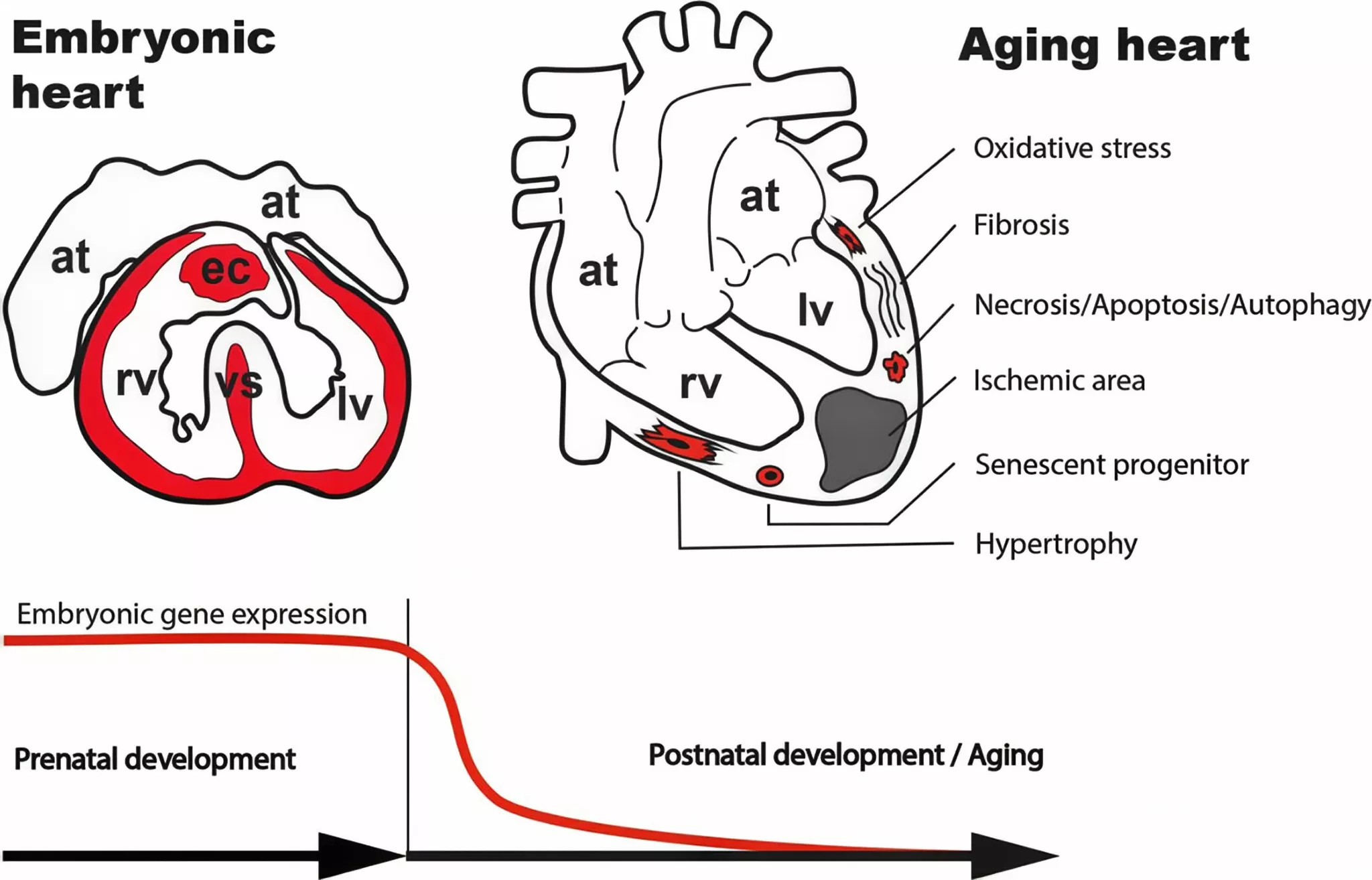
Figure 2 Alterations in the embryonic gene expression program, combined with frequent exposure to external and internal stress factors, initiate various pathological alterations in the adult heart throughout our postnatal life.
Stabilizing the structure of new blood vessels
In addition to inducing angiogenesis, TB 500 plays a crucial role in stabilizing the structure of new blood vessels. It regulates the composition and remodeling of the extracellular matrix (ECM), enabling new blood vessels to integrate more effectively into surrounding tissues. TB 500 promotes the synthesis of ECM components such as collagen and fibronectin while balancing matrix metalloproteinases (MMPs) and their tissue inhibitors (TIMPs), preventing new blood vessels from being damaged by excessive degradation. In the later stages of angiogenesis, this action of TB 500 helps maintain the integrity and function of new blood vessels, ensuring that injured muscles continue to receive adequate nutrient supply and promoting muscle recovery.
(3) Anti-inflammatory and Immune Regulatory Mechanisms
Inhibition of Inflammatory Factor Expression
Muscle injury triggers an inflammatory response, and excessive inflammation can cause further damage to muscle tissue. TB 500 possesses anti-inflammatory properties, inhibiting the expression of various inflammatory factors such as tumor necrosis factor-α (TNF-α), interleukin-1β (IL-1β), and interleukin-6 (IL-6). During inflammation, TB 500 can bind to receptors on the surface of immune cells, activate intracellular anti-inflammatory signaling pathways, and inhibit the transcription and translation of inflammatory cytokine genes. In animal inflammation models, after using TB 500, the levels of TNF-α, IL-1β, and IL-6 in muscle tissue were significantly reduced, and the infiltration of inflammatory cells was also decreased. This indicates that TB 500 alleviates the inflammatory response following muscle injury by inhibiting the expression of inflammatory factors, thereby promoting muscle recovery.
Regulating immune cell function
TB 500 can also regulate the function of immune cells, promoting immune responses that favor tissue repair. It can influence the polarization of macrophages, causing them to transition from pro-inflammatory M1 type to anti-inflammatory and pro-repair M2 type. M2-type macrophages can secrete various growth factors and cytokines, such as insulin-like growth factor-1 (IGF-1) and transforming growth factor-β (TGF-β), which help promote muscle cell proliferation and repair. Additionally, TB 500 can regulate T lymphocyte function, suppressing excessive immune responses to prevent damage to the body’s own muscle tissue. By modulating immune cell function, TB 500 coordinates the immune response following muscle injury, thereby promoting muscle repair and recovery.
The Role of TB 500 in Muscle Recovery
(1) Accelerating Muscle Injury Repair
Shortening Repair Time
Whether it is acute muscle strain or chronic muscle fatigue, TB 500 can significantly shorten the repair time of muscle damage. In animal experiments, after causing muscle damage, the experimental group treated with TB 500 showed a significantly accelerated healing rate at the site of muscle damage. Histological analysis revealed that the experimental group exhibited more new muscle fibers and blood vessels in a shorter time, and inflammatory cell infiltration also subsided more rapidly. In human studies, for athletes with muscle strains caused by high-intensity training, after local injection or systemic administration of TB 500, patients reported earlier relief of muscle pain and a significant reduction in the time required for muscle function recovery, enabling them to return to training and competition more quickly.
Improving repair quality
TB 500 not only accelerates the repair speed of muscle injuries but also improves the quality of repair. Through mechanisms such as promoting myoblast migration, satellite cell activation and differentiation, and angiogenesis, TB 500 makes the repaired muscle tissue structurally and functionally closer to normal muscle. Repaired muscle fibers are arranged more uniformly, and muscle contractile force and endurance are better restored. After using TB 500 to repair injured muscles, the differences in maximum contractile force and fatigue tolerance time between repaired and uninjured muscles are smaller, indicating that TB 500 effectively improves the quality of muscle injury repair and reduces the long-term impact of muscle injury on motor function.
(2) Alleviating Muscle Fatigue
Energy Metabolism Regulation
Muscle fatigue is closely related to energy metabolism disorders. TB 500 can regulate energy metabolism processes within muscle cells, improving energy supply efficiency and thereby alleviating muscle fatigue. It promotes mitochondrial biogenesis and function, increasing mitochondrial number and activity, enabling muscle cells to perform aerobic respiration more efficiently and produce more adenosine triphosphate (ATP). Additionally, TB 500 can regulate the activity of enzymes involved in glucose metabolism and fatty acid metabolism, optimizing the utilization of energy substrates. During exercise, TB 500 can promote the oxidative breakdown of fatty acids, providing muscles with more energy, reducing lactic acid accumulation, and delaying the onset of muscle fatigue.
Oxidative Stress Regulation
During exercise, a large amount of reactive oxygen species (ROS) is produced, leading to oxidative stress, which is one of the primary causes of muscle fatigue. TB 500 has antioxidant properties, enhancing the activity of intracellular antioxidant enzymes such as superoxide dismutase (SOD) and glutathione peroxidase (GPx), reducing ROS accumulation, and mitigating oxidative stress-induced damage to muscle cells. In animal experiments, animals pretreated with TB 500 exhibited significantly lower oxidative stress markers in muscle tissue and milder muscle fatigue compared to the control group after prolonged exercise. This study demonstrates that TB 500 alleviates muscle cell damage and reduces muscle fatigue by regulating oxidative stress responses, thereby maintaining normal muscle function.
(3) Promoting Muscle Growth and Development
Stimulating Muscle Protein Synthesis
TB 500 promotes muscle protein synthesis by activating the mTOR signaling pathway within cells. mTOR is a key molecule regulating protein synthesis, promoting ribosomal biosynthesis and the activity of protein translation initiation factors, thereby increasing the rate of muscle protein synthesis. TB 500 also regulates the expression of transcription factors, including NF-κB, which indirectly influences muscle protein synthesis. In vitro cell culture experiments showed that adding TB 500 significantly increased protein synthesis in muscle cells. In animal experiments, animals treated with TB 500 over the long term exhibited significant increases in muscle mass and strength. TB 500 promotes muscle growth and development by stimulating muscle protein synthesis.
Regulating muscle fiber type conversion
Muscle fiber types are divided into fast-twitch and slow-twitch fibers, which differ in exercise performance and metabolic characteristics. TB 500 can regulate muscle fiber type conversion, directing it toward a direction more conducive to enhancing athletic performance. Research has shown that TB 500 promotes the expression of slow-twitch fiber-related genes, such as the slow-type myosin heavy chain (MyHC) gene, while inhibiting the expression of fast-twitch fiber-related genes.
Application of TB 500 in Muscle Recovery
(1) Application in Sports
Athlete Injury Prevention and Recovery
In competitive sports, athletes face high-intensity training and competitions, resulting in a high incidence of muscle injuries. By administering TB 500 appropriately before training, muscle resistance to injury can be enhanced, reducing the risk of injuries such as muscle strains and sprains. When athletes sustain muscle injuries, timely use of TB 500 for treatment can accelerate injury repair, shorten recovery time, and enable athletes to return to competition more quickly. In some track and field and football events, athletes have begun to experiment with TB 500 to promote muscle injury recovery and have achieved good results.
Enhancing Athletic Performance
TB 500 can also enhance athletic performance by alleviating muscle fatigue and promoting muscle growth and development. In endurance sports, TB 500’s regulatory effects on energy metabolism and oxidative stress can delay the onset of muscle fatigue, thereby improving athletes’ endurance levels. In strength sports, TB 500’s stimulation of muscle protein synthesis and regulation of muscle fiber type conversion can increase muscle strength and explosive power.
(2) Applications in Rehabilitation Medicine
Muscle Injury Rehabilitation Treatment
In rehabilitation medicine, TB 500 can be used as an adjunctive therapy for muscle injury rehabilitation. For patients with muscle injuries caused by trauma, surgery, or other factors, combining conventional rehabilitation therapy with TB 500 use can enhance rehabilitation outcomes. In patients with muscle injuries following fractures, local injection or systemic administration of TB 500 alongside physical therapy and rehabilitation training can accelerate the recovery of muscle strength and function, thereby shortening the rehabilitation period. Rehabilitation physicians can tailor TB 500 treatment plans based on the patient’s specific condition, including dosage, frequency, and route of administration, to achieve optimal rehabilitation outcomes.
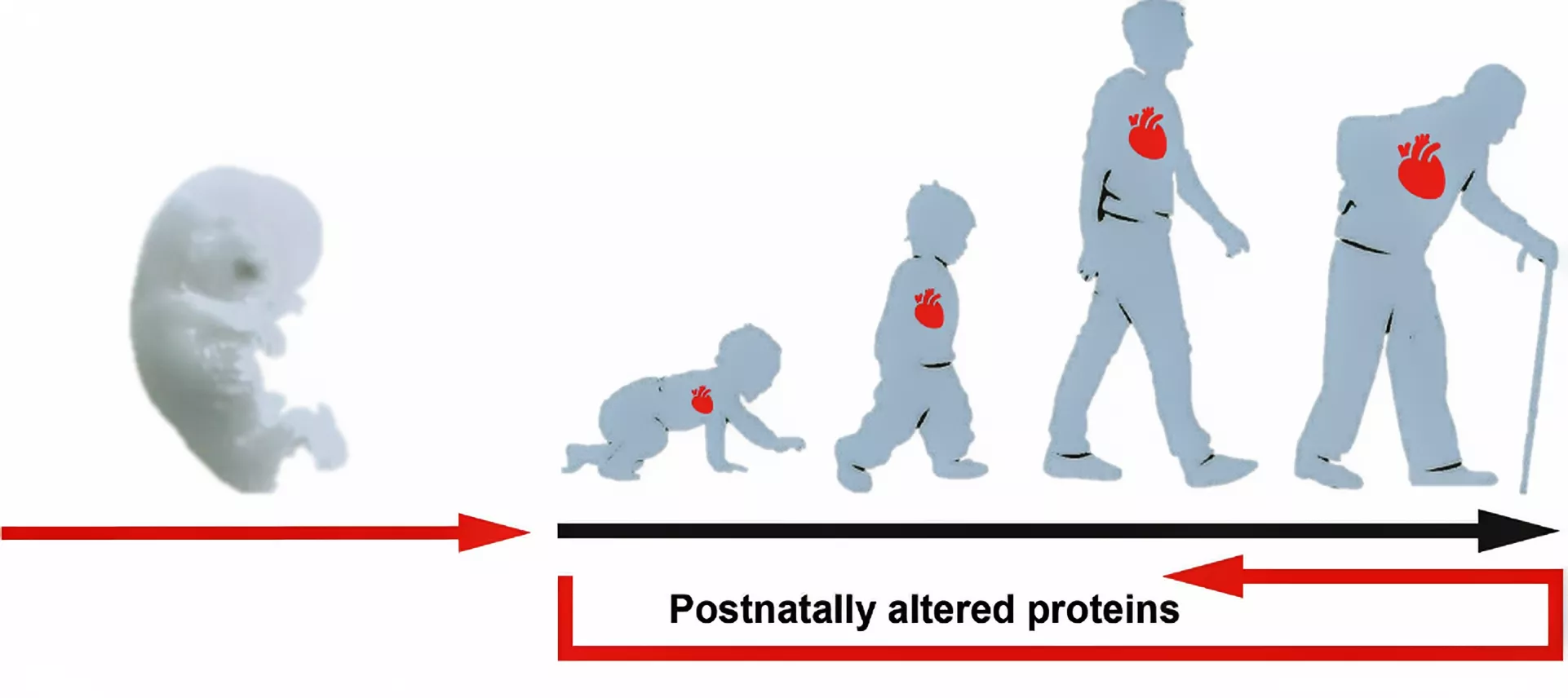
Figure 3: Concept of utilizing prenatally active secreted molecules to restore organ function in the elderly.
Treatment of muscle atrophy associated with neurological disorders
Certain neurological diseases, such as spinal cord injury and amyotrophic lateral sclerosis (ALS), can lead to muscle atrophy. TB 500 can exert a therapeutic effect on muscle atrophy associated with neurological diseases by promoting muscle cell proliferation and inhibiting muscle cell apoptosis. In animal experiments, for spinal cord injury-induced muscle atrophy models, the use of TB 500 resulted in a significant improvement in the degree of muscle atrophy, with muscle mass and strength partially restored.
Conclusion
TB 500 can promote muscle recovery. Through mechanisms such as cell migration and regeneration, angiogenesis, and anti-inflammatory and immune regulation, it plays an important role in muscle injury repair, fatigue relief, and growth and development. In the field of sports, it can help athletes prevent and treat muscle injuries and improve athletic performance; in the field of rehabilitation medicine, it can be used for muscle injury rehabilitation treatment and the treatment of muscle atrophy associated with neurological diseases.
Sources
[1] Bo Z, Mingjun L. Recovery from muscle injuries after high-intensity training in table tennis[J]. Revista Brasileira De Medicina Do Esporte, 2023,29.DOI:10.1590/1517-8692202329012022_0550.
[2] Zhang D, Fan G. Application of conjugated materials in muscle movement recovery process[J]. Frontiers in Chemistry, 2023,11:1246926.DOI:10.3389/fchem.2023.1246926.
[3] Roveratti M C, Jacinto J L, Oliveira D B, et al. Effects of beta-alanine supplementation on muscle function during recovery from resistance exercise in young adults[J]. Amino Acids, 2019,51(4):589-597.DOI:10.1007/s00726-018-02686-y.
[4] Ferreira D V, Gentil P, Soares S, et al. Recovery of pectoralis major and triceps brachii after bench press exercise[J]. Muscle & Nerve, 2017,56(5):963-967.DOI:10.1002/mus.25541.
[5] Markos F, Campion D P, Carey M, et al. An investigation into the mechanism of action of almitrine on isolated rat diaphragm muscle fatigue[J]. Respiration, 2002,69(4):339-343.DOI:10.1159/000063267.
[6] Noda K. Mechanism of the restoration of intracellular cation level in the muscle immersed with sucrose solution[J]. The Kurume Medical Journal, 1965,12:119-129.
[7] Bock-Marquette I, Maar K, Maar S, et al. Thymosin beta-4 denotes new directions towards developing prosperous anti-aging regenerative therapies[J]. International Immunopharmacology, 2023,116:109741.DOI:https://doi.org/10.1016/j.intimp.2023.109741.
Product available for research use only:
<
By Peplyte 1 month ago
ALL ARTICLES AND PRODUCT INFORMATION PROVIDED ON THIS WEBSITE ARE SOLELY FOR INFORMATION DISSEMINATION AND EDUCATIONAL PURPOSES.
The products provided on this website are intended exclusively for in vitro research. In vitro research (Latin: *in glass*, meaning in glassware) is conducted outside the human body. These products are not pharmaceuticals, have not been approved by the U.S. Food and Drug Administration (FDA), and must not be used to prevent, treat, or cure any medical condition, disease, or ailment. It is strictly prohibited by law to introduce these products into the human or animal body in any form.
Dermorphin is a potent opioid peptide isolated from the skin of certain frog species.
The Mechanism of Dermorphin’s Analgesic Effects
Interaction with Opioid Receptors
Dermorphin primarily exerts its analgesic effects by interacting with the μ-opioid receptor (MOP). The μ-opioid receptor is a key member of the opioid receptor family and plays a crucial role in pain modulation. Dermorphin and its analogues can specifically bind to the MOP receptor, akin to a key fitting into a lock, thereby activating downstream signaling pathways. [Cys (ATTO 488) 8] Dermorphin – NH₂ (DermATTO488), a novel fluorescent Dermorphin analog based on ATTO488, binds to HEK and CHO cells expressing human MOP receptors and exhibits similar binding characteristics to Dermorphin. It stimulates G protein binding, promotes GTPγ[35S] binding, thereby activating related signal transduction, and ultimately inhibits pain signal transmission. Dermorphin also stimulates ERK1/2 phosphorylation, further influencing intracellular biological processes and participating in the regulation of analgesic effects.
Inhibition of pain signal transmission at the spinal cord level
Experimental studies have shown that Dermorphin significantly inhibits pain signal transmission at the spinal cord level. In experiments, intravenous infusion of 0.16 mg/kg Dermorphin induced a significant and sustained increase in the nociceptive flexion reflex threshold in healthy volunteers, and this effect was equally pronounced in subjects with complete chronic spinal cord injury, indicating that Dermorphin primarily inhibits nociceptive transmission at the spinal cord level. This mechanism of action may be related to Dermorphin’s regulation of neurotransmitter release within the spinal cord or its direct action on spinal cord neurons. The spinal cord serves as a critical hub for pain signal transmission, and Dermorphin’s modulation of its function effectively blocks the upward transmission of pain signals to the brain, thereby achieving analgesic effects.
Unique receptor action characteristics
Although naloxone can completely antagonize the effects of morphine and opioid analogs, it can only partially (approximately 50%) reverse Dermorphin’s inhibitory effect on nociceptive spinal reflexes. This suggests that Dermorphin interacts with different groups of spinal opioid receptors when inducing analgesia, and its mechanism of action differs from that of traditional opioid drugs. This mode of action confers certain advantages in analgesic applications, such as reducing common side effects associated with traditional opioid drugs.
Analgesic Performance of Dermorphin in Animal Experiments
Effective Analgesia in Multiple Animal Models
In various animal pain models, Dermorphin demonstrated potent analgesic effects. In experiments such as tail-flick, hot plate, tail pinch, formalin, and acetic acid writhing tests, intraperitoneal administration of Dermorphin and its related peptides reduced animals’ sensitivity to pain and increased their pain thresholds. These experiments simulated pain of varying degrees and types, comprehensively validating Dermorphin’s analgesic capabilities. In the hot plate test, after administering Dermorphin to animals, the latency period for licking their paws or jumping was significantly prolonged, indicating a weakened pain response to thermal stimulation, thereby demonstrating Dermorphin’s excellent analgesic effect.
Comparative Advantages Over Other Analgesic Drugs
Compared to traditional analgesic drugs such as morphine, Dermorphin exhibits significant advantages in certain aspects. In animal models, after intracerebroventricular administration, Dermorphin exhibited more selective and potent analgesic effects than morphine, with a longer duration of action. Some synthesized Dermorphin tetrapepetides also demonstrated high analgesic activity, with analgesic potency approximately 1,500 times and 17 times that of morphine after intracerebroventricular or subcutaneous administration in mice, respectively. These advantages offer promising prospects for the clinical application of Dermorphin in analgesia, potentially addressing shortcomings of traditional analgesic drugs, such as insufficient analgesic efficacy or duration.
Research and Application Exploration of Dermorphin in Humans
Early Clinical Trial Results
In 1985, a randomized, placebo-controlled clinical trial on the use of Dermorphin for postoperative pain demonstrated that Dermorphin administered via intrathecal injection significantly outperformed placebo and morphine (used as a reference compound) in terms of analgesic efficacy. This landmark study provided crucial supporting evidence for the clinical application of Dermorphin in analgesia, highlighting its immense potential in postoperative pain management.
Potential Application Scenarios
Postoperative Pain Management: Surgical trauma can cause severe postoperative pain, significantly impacting patients’ recovery and quality of life. Dermorphin’s demonstrated efficacy in alleviating postoperative pain in early clinical trials positions it as a promising new option for postoperative analgesia. Compared to traditional postoperative analgesic drugs, its unique mechanism of action and advantages—such as reduced risk of drug tolerance and dependence—offer patients a safer and more effective analgesic solution.
Chronic pain treatment: For patients with chronic pain, especially those who have poor responses to traditional analgesic drugs or experience significant side effects, Dermorphin may serve as a valuable alternative treatment option. In the management of chronic pain in cancer patients, current treatment methods still have certain limitations. The emergence of Dermorphin brings new hope to this patient population. Its long-lasting analgesic effects can better control persistent pain in cancer patients and improve their quality of life.
Palliative care: In palliative care settings, alleviating patients’ pain is the top priority. Dermorphin’s potent analgesic properties make it a promising candidate for alleviating end-of-life pain in palliative care. Through appropriate administration methods and dose adjustments, it can help patients reduce suffering and enhance comfort during the final stages of life.
Conclusion
As a substance with a unique analgesic mechanism and significant analgesic effects, Dermorphin holds promising applications in the field of pain management.
Sources
[1] Giakomidi D, Bird M F, McDonald J, et al. Evaluation of [Cys(ATTO 488)8]Dermorphin-NH2 as a novel tool for the study of μ-opioid peptide receptors[J]. Plos One, 2021,16(4):e250011.DOI:10.1371/journal.pone.0250011.
[2] Hesselink J, Schatman M E. Rediscovery of old drugs: the forgotten case of dermorphin for postoperative pain and palliation[J]. Journal of Pain Research, 2018,11:2991-2995.DOI:10.2147/JPR.S186082.
[3] Guzevatykh L S, Voronina T A, Emel’Ianova T G, et al. Comparative analysis of analgesic activities of dermorphin, [DPro6]-dermorphin, and their C-terminal tripeptides[J]. Izv Akad Nauk Ser Biol, 2007(5):577-582. http://www.ncbi.nlm.nih.gov/entrez/query.fcgi?cmd=Retrieve&db=pubmed&dopt=Abstract&list_uids=18038625&query_hl=1
[4] Sandrini G, Degli U E, Salvadori S, et al. Dermorphin inhibits spinal nociceptive flexion reflex in humans[J]. Brain Research, 1986,371(2):364-367.DOI:10.1016/0006-8993(86)90376-8.
[5] Salvadori S, Marastoni M, Balboni G, et al. Synthesis and opioid activity of dermorphin tetrapeptides bearing D-methionine S-oxide at position 2[J]. Journal of Medicinal Chemistry, 1986,29(6):889-894.DOI:10.1021/jm00156a003.
Product available for research use only: